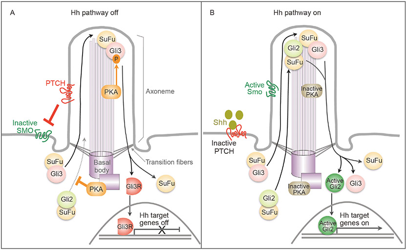
Figure 1.
Overview of Hh signal transduction. Vertebrate Hh signaling relies on protein trafficking at the primary cilium, a centrosome organized, microtubule-based cell surface organelle. The membrane of the cilium is continuous with the cytoplasmic membrane, but the ciliary cytosol is separated from the cytoplasm via transition fibers that function as filter for material exchange between ciliary axoneme and the cytoplasm. A, When the Hh pathway is off, Ptch resides in the cilium and inhibits Smo. Active PKA phosphorylates Gli3, resulting in Gli3’s proteolytical processing into Gli3R, a transcription repressor. Active PKA also phosphorylates Gli2 and inhibits its trafficking to the cilium tip, preventing its activation. Note that both Gli proteins complex with SuFu, a negative regulator of the Hh pathway. B, Upon binding to the ligand Shh, Ptch exits from the cilium, and Smo is activated and accumulated in the cilium. PKA is inactivated and stops phosphorylating Gli proteins. As a result, both Gli proteins dissociate from SuFu. Gli3 remains in the full-length form and Gli2 is activated. Active Gli2 enters the nucleus to turn on the transcription of Hh target genes.

