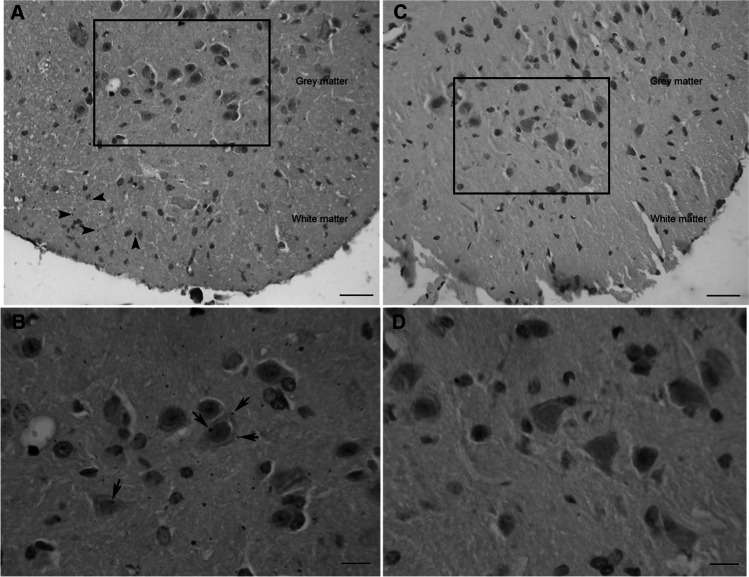
Fig. 2.
Mercury accumulation at a lumbar level in the spinal cord of chronically exposed mice (A–B) and animals from the control site (C–D). A AMG staining in white matter (arrowheads) and grey matter; scale bar: 25 μm. B Magnified microphotography from the squared area in A. Mercury deposit could be observed adhered to the membrane of different motor neurons (arrows); scale bar: 10 μm. C Lumbar segment of the spinal cord of a mouse from Rabo de Peixe. Note that there is no AMG staining in both grey and white matter; scale bar: 25 μm. D Inset from the squared zone in C where no mercury deposits are present in the grey matter of rodents from de control site; scale bar: 10 μm

