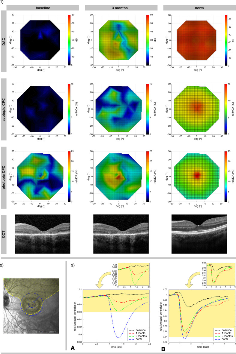
Figure 1.
Illustration of treatment effects of P3, the youngest subject who responded well to the therapy. 1) Retinotopy of the photoreceptor rescue, measured via DAC cyan (upper row; the color-coded bar represents the threshold decrease from 0 dB), scotopic CPC and photopic CPC (the second and third row, the color-coded bar represents the relMCA value in %). Foveal horizontal lines of the OCT images from the same visits are shown in the bottom line. The findings are presented in comparison to a normative measurement in healthy eyes (right column). 2) The extent of the surgically induced subretinal bleb for the administration of voretigene neparvovec in the left eye of P3 is outlined in yellow, with an overlay of the OCT grid used for the calculations of the retinal volumes (inner circle: approximately 3 degrees macular region; outer circle: 12 degrees macular region, further subdivided into the four quadrants of the OCT retinal volume analysis). 3) Averaged relative pupil response in the 15 degrees macular area of P3 for scotopic (A) and photopic (B) stimuli over time. At baseline (black lines) there was no measurable response of the pupil to scotopic stimuli and a decreased pupil reaction for photopic stimuli. The improvement at 1 month (red lines) increased further at 3 months (green lines) for scotopic response and remained stable at near to normal values for photopic response. Lower diagrams show the responses for both scotopic and photopic stimuli in relation to normative responses of healthy eyes (blue lines). CPC, chromatic pupil campimetry; DAC, dark-adapted chromatic perimeter; OCT, optical coherence tomography; relMCA, relative maximal constriction amplitude.

