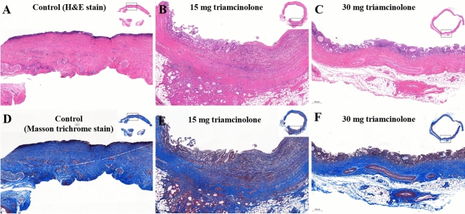
Figure 3.
Microscopic pathology. Microscopic appearance of H&E-stained and Masson trichrome-stained sections of the bile duct 4 weeks after stent insertion. Control group (A) showing eroded epithelium, thick fibrosis, and moderate inflammation. Triamcinolone (15 mg) steroid-eluting stent group (B) showing residual erosion with regenerated epithelium, moderate inflammation, and partially resolved fibrosis. Triamcinolone (30 mg) steroid-eluting stent group (C) showing complete epithelial recovery, mild to moderate inflammation, and less fibrosis than the other groups.

