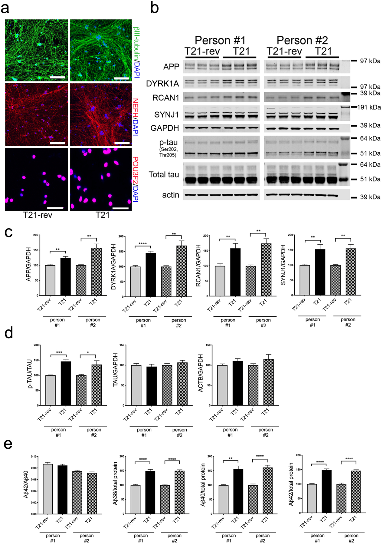
Figure 1. Trisomy 21 neurons express elevated phosphorylated tau and higher Aβ levels.

Pairs of T21 and T21-rev iPSC lines derived from two individuals were differentiated to neuronal fate using the NGN2-direct induction protocol and analyzed at d21.
a) Representative immunostaining for neuronal markers. Scale bars for top four panels = 200 μm, for bottom two panels = 50 μm. b) Representative WBs showing protein levels of chromosome 21 encoded proteins, tau and loading controls. c,d) WB quantification of protein levels encoding chromosome 21 genes (c) and of p-tau, tau, actin, and GAPDH (d). e) Measurements of Aβ in the media (MSD 6E10 ELISA kit). In (c-e), for each condition, three differentiations were performed and analyzed with 3 wells per differentiation. Data were normalized to the T21-rev for each differentiation round. Two-tailed, unpaired t-test with Welch correction; *p<0.05; **p<0.01; ***p<0.005; ****p<0.001.
