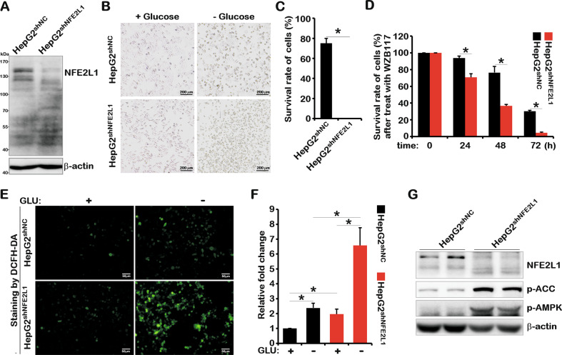
Fig. 1. NFE2L1 knockdown causes HepG2 cells to be more sensitive to glucose starvation.
A The total protein of HepG2shNC and HepG2shNFE2L1 cells were collected, and then, the expression of NFE2L1 and β-actin were detected by western blotting (WB). The HepG2shNC and HepG2shNFE2L1 cells were cultured in DMEM with or without glucose (2 g/L or 0 g/L) for 18 h, the morphology of cells was observed under a microscope (B), and the survival rate of cells was detected by a CCK8 kit (C). D The HepG2shNC and HepG2shNFE2L1 cells were treated with WZB117 (200 μM), and the survival rate of cells was detected at 0, 24, 48, and 72 h using a CCK8 kit. The HepG2shNC and HepG2shNFE2L1 cells were cultured in DMEM without glucose for 6 h, and then, the reactive oxygen species (ROS) in cells were staining with DCFH-DA (10 μM) for 20 min and imaged by microscopy (E); the intensity of fluorescence was quantitated (F). The scale is 50 μm. G The total protein of HepG2shNC and HepG2shNFE2L1 cells were collected, and the expression of NFE2L1, pAMPK, pACC, and β-actin were detected by WB. n ≥ 3, *p < 0.05.

