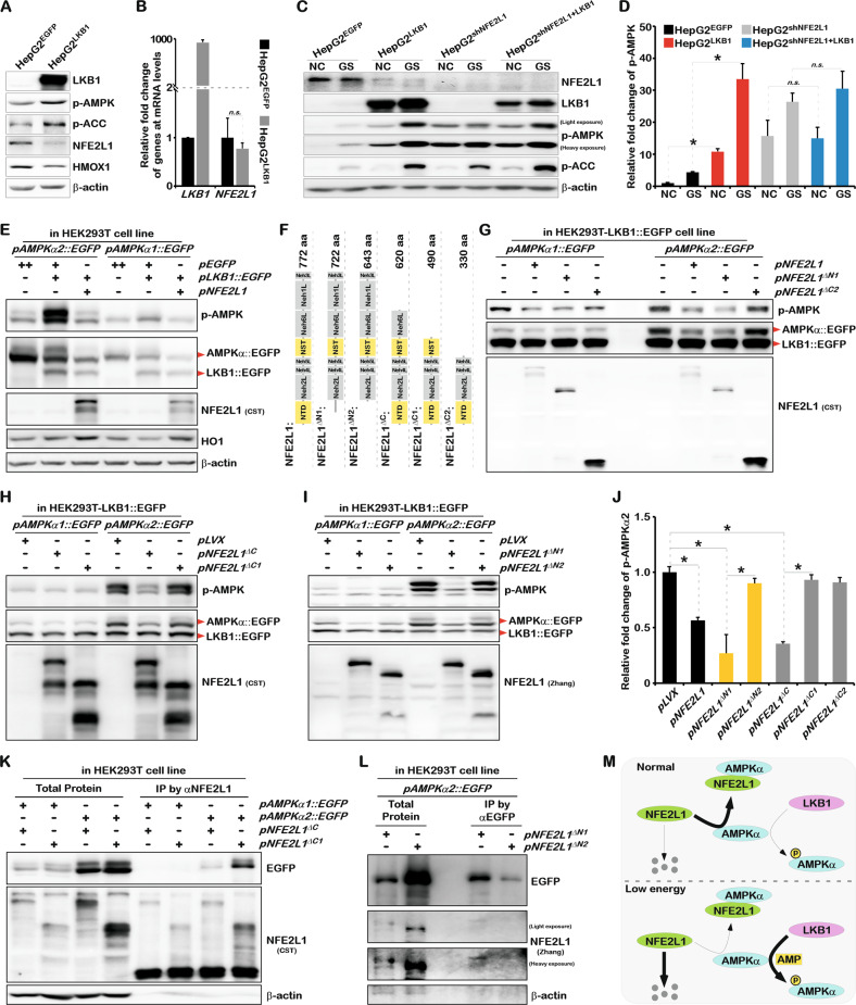
Fig. 6. NFE2L1 disrupts the phosphorylation of AMPK mediated by LKB1 by directly interacting with AMPK.
A The expression of LKB1, p-AMPK, p-ACC, NFE2L1, HO1, and β-actin in HepG2EGFP and HepG2LKB1 cells were detected by WB. HepG2EGFP and HepG2LKB1 cells were constructed via lentiviral infection. B The expression of LKB1 and NFE2L1 in HepG2EGFP and HepG2LKB1 cells was detected by qPCR, with β-actin used as the internal control. C HepG2EGFP, HepG2LKB1, HepG2shNC, and HepG2shNFE2L1+LKB1 cells were treated with glucose starvation (GS) for 4 h, and the expression of NFE2L1, LKB1, p-AMPK, p-ACC, and β-actin were detected by WB. D Relative fold-changes in p-AMPK in (C). E The pAMPKα1::EGFP and pAMPKα2::EGFP were co-transfected with pEGFP, pLKB1::EGFP, pNFE2L1 plasmids into HEK293T cells, respectively. And the expression of p-AMPK, EGFP, NFE2L1, LKB1, HO1, and β-actin were detected by WB. F Schematic diagram of NFE2L1 truncated protein. The pAMPKα1::EGFP, pAMPKα2::EGFP were co-transfected with pNFE2L1, pNFE2L1ΔN1, pNFE2L1ΔC2 (G) or pLVX, pNFE2L1ΔC, pNFE2L1ΔC1 (H) or pLVX, pNFE2L1ΔN1, pNFE2L1ΔN2 (I) into HEK293T-LKB1::EGFP cells, respectively. And the expression of p-AMPK, EGFP, and NFE2L1 were detected by WB. J Statistical analysis of the changes of p-AMPKα2 in (G–I). K The pAMPKα1::EGFP and pAMPKα2::EGFP were co-transfected with pNFE2L1ΔC and pNFE2L1ΔC1 plasmids into HEK293T cells, respectively. And the total protein was collected with non-denaturing lysate buffer after 48 h. The specific antibody for NFE2L1was used for Co-IP experiments; then EGFP, NFE2L1 and β-actin were detected by WB. L The pAMPKα2::EGFP, pNFE2L1ΔN1, pNFE2L1ΔN2 plasmids were co-transfected into HEK293T cells, and the total protein was collected with non-denaturing lysate buffer after 48 h. The specific antibody for EGFP was used for Co-IP experiments; then EGFP, NFE2L1, and β-actin were detected by WB. M The model diagram of NFE2L1 involved in regulating AMPK signaling. n ≥ 3, *p < 0.05.

