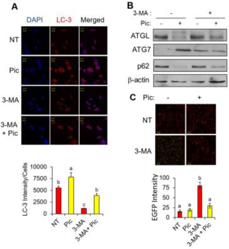
Fig. 3. Piceatannol treatment stimulates autophagy in vitro.
(A) Immunofluorescence analysis of Hela cells treated with 50 μM piceatannol (Pic), 5 mM 3-MA or the combination of Pic and 3-MA in serum-free medium for 2 hr (LC3: red and DAPI: blue) (top). Quantification of the fluorescence intensity of LC3 was shown (bottom). (B) Hela cells described in panel A were then subjected to Western blot analysis for ATGL, ATG7, p62 and β-actin. (C) 3T3-L1 adipocytes were treated with piceatannol (25 μM) for 90 min followed by GFP-expressing S. typhimurium infection for 30 min. α-tubulin (red) and EGFP (green) signals were visualized by confocal microscopy (top). Quantification of the fluorescence intensity of EGFP was shown (bottom). Scale bar indicates 20 μm. Data are presented as means ± SEM and different letters indicate statistical significance. NT: non-treated.

