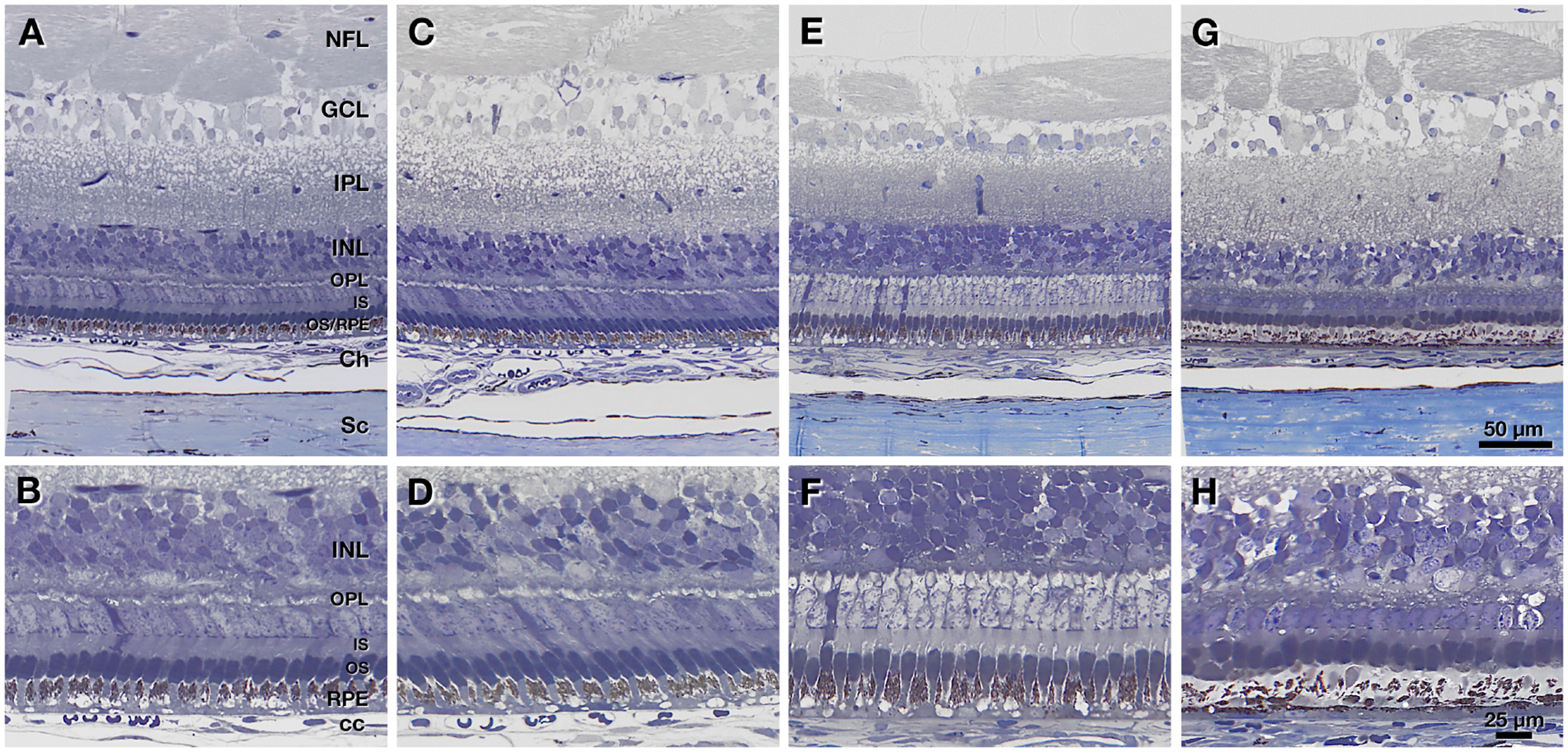
Figure 8:

Histology of control (untreated, n=1), sham treated (FD + 5 × sham, n=1) and genipin treated (FD + 5 × 20 mM GEN, n=2) tree shrew eyes. A, C, E, and G show overviews of retina and choroid from the peripapillary region close to the ONH. B, D, F, and H show high-magnification views of outer retina at the same locations. The ganglion cell layer (GCL) was poorly preserved and highly vacuolated in all specimens. A, B: Normal eye has thick inner retinal layers with vertically aligned photoreceptors and apical processes of the retinal pigment epithelium (RPE), the latter recognized by the content of darkly pigmented melanosomes. C, D: Sham-injected eye also has thick inner retinal layers with organized vertically aligned photoreceptors. E, F: An eye receiving the highest genipin dose exhibits inner retinal thinning. Photoreceptors are still organized and vertically aligned. Pale staining of photoreceptor cell bodies in the outer nuclear layer could not be definitively attributed to an effect of genipin, an effect of the fixation, or both. G, H: Another eye receiving the highest dose of genipin exhibits retinal thinning and noticeable degeneration of photoreceptors and RPE, manifest as outer nuclear layer thinning, shortening and absence of outer segments, and reduction in the area of pigmented apical processes. Abbreviations: NFL, nerve fiber layer; GCL, ganglion cell layer; IPL, inner plexiform layer; INL, inner nuclear layer; OPL, outer plexiform layer; IS, inner segments of photoreceptor; OS, outer segments of photoreceptors; RPE, retinal pigment epithelium; Ch, choroid; CC, choriocapillaris; Sc, sclera.
