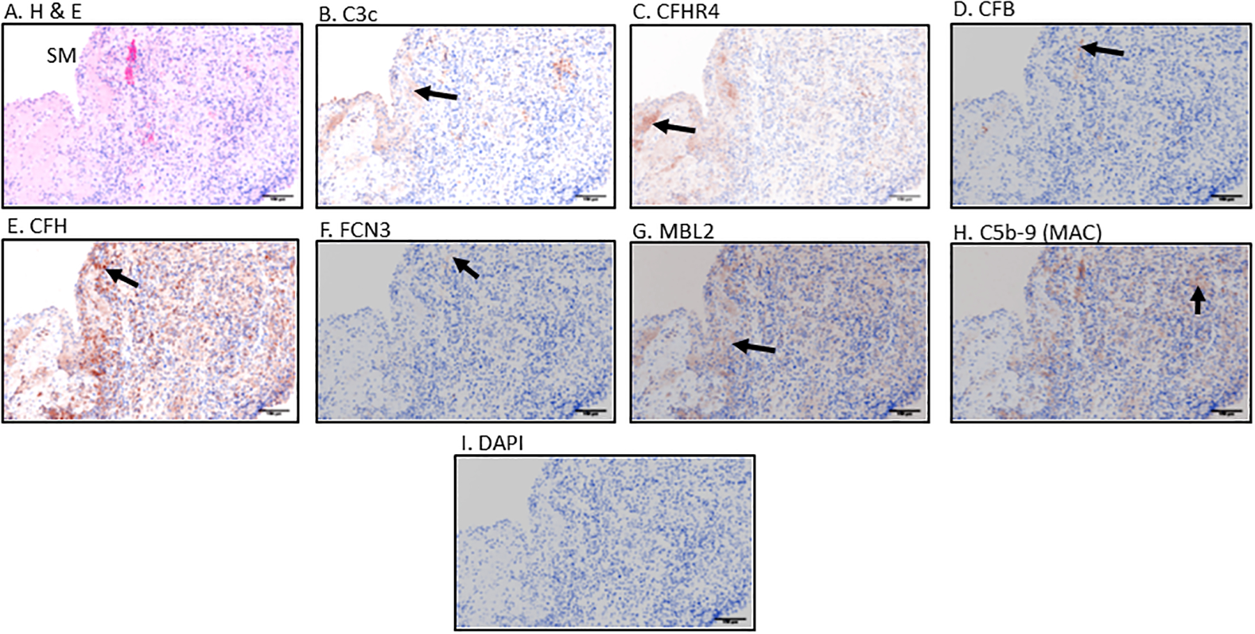
Figure 4.

Immunohistochemical (IHC) staining, standardization and examination of Early RA synovial biopsy for seven complement proteins including Hematoxylin and Eosin (H &E). Top panel from left to right A. H & E. B. C3c C. CFHR4 D. CFB; Center panel from left to right E. CFH F. FCN3 G. MBL2 H. C5b-9 (MAC) and bottom panel I. DAPI. Positive brown color staining for each complement protein has been indicated by a black arrow. IHC staining for each complement marker was repeated two times using different human tissue. Magnification =20x, Scale bar = 100μm
