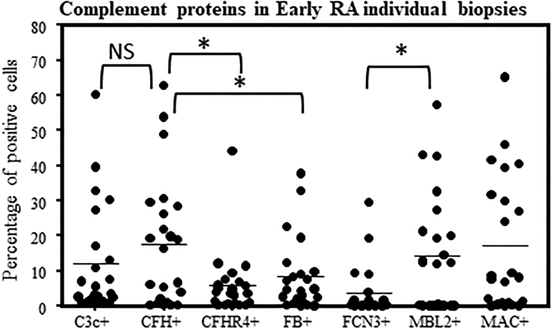
Figure 5.

Examples of complement protein positive cells in the synovial biopsies from Early RA patients. Quantitative data for seven complement protein positivity in each synovial biopsy were generated by scanning multiple areas as described in the Methods. Percentage of positive cells for various complement proteins in Early RA synovial biopsies. MIHC quantitative data were obtained using a single synovial biopsy and repeated with 1–2 sections from each patient. Mean percentage and variations in percentage of complement protein positive cells in individual early RA synovial biopsies (n = 23). *p < 0.05 considered significant.
