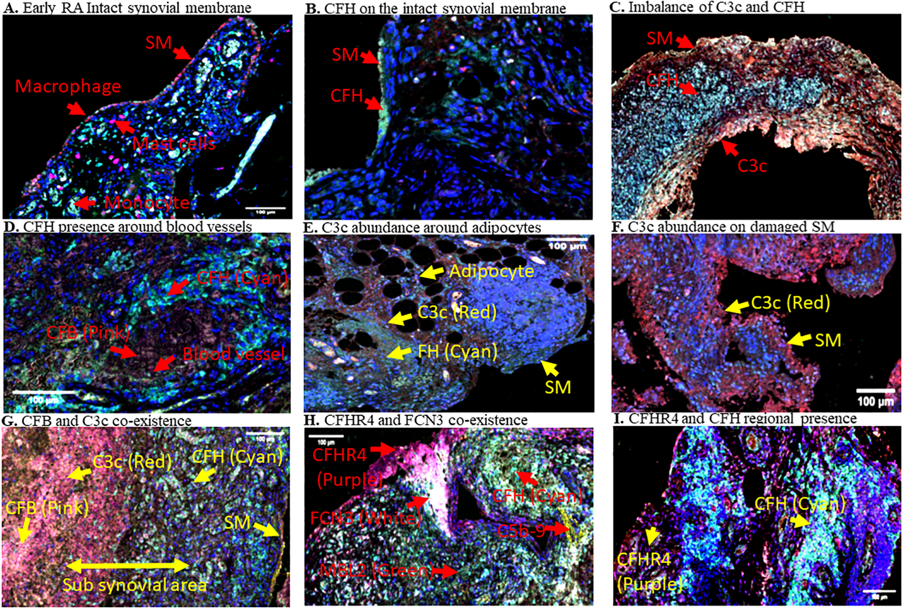
Figure 7.

Representative MIHC composite images showing presence of immune cells and regional imbalance of complement proteins on the synovial membrane and sub-synovial areas in Early RA biopsies. A. Macrophages (red color) are predominately present on the single cell synovial lining at early stage of the disease. B. CFH presence was found predominantly in the vicinity of synovial membrane (SM) cells in some biopsies in Early RA. C. Imbalance of CFH and C3c localization in some regions of biopsies. D. Presence of CFH around large blood vessles. E. C3c localization between and around adipocytes. F. C3c presence on damaged SM. G. CFB and C3c co-existence away from CFH in deep subsynovial liming area in Early RA. H. CFHR4 and FCN3 co-localization in the SM and subsynovial lining area. I. CFHR4 and CFH imbalance in the SM and sub-synovial lining area. Various complement proteins, and the SM have been indicated as marked by a yellow arrow in each biopsy. Presence of individual complement protein in each biopsy composite image has been shown by using a false color coding key i.e. CFH (cyan), C3c (red), CFHR4 (purple), CFB (pink), FCN3 (white), and C5b-9 (Mac)(yellow). These experiments were repeated two times using a single biopsy from each patient with 1–2 sections on each patient. Representative photos are from Early RA synovial biopsies obtained from unique individuals (n = 9). Scale bar = 100μm
