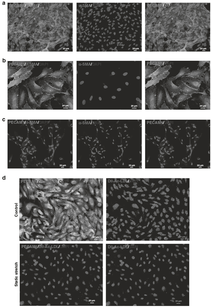
Fig. 1. Static stretch triggers EndMT in isolated endothelial cells.

a HCAEC present with cobblestone morphology typical for endothelial cells. b, c Representative histological sections of control endothelial cells and stretched endothelial cells are shown. Both slides were stained with the EC marker, PECAM1 (green), the mesenchymal marker, αSMA (red), and nuclei in blue (DAPI). Endothelial cells in the control picture only stained for the EC marker PECAM1 indicative of the endothelial phenotype. In comparison, stretched endothelial cells were positive for both markers. Double-staining with an EC and a mesenchymal marker is indicative of active EndMT. d To confirm the results of stretch-induced EndMT in HCAEC, we added the functional endothelial marker Dil complex acetylated Low Density Lipoprotein (Dil-Ac-LDL) to the media of the cell culture dish. Dil-Ac-LDL (red) is only internalized by endothelial cells and fluoresces upon uptake. As indicated by the representative pictures, stretched cells had no functional endothelial cells compared to the endothelial cells (green) in the control group.
