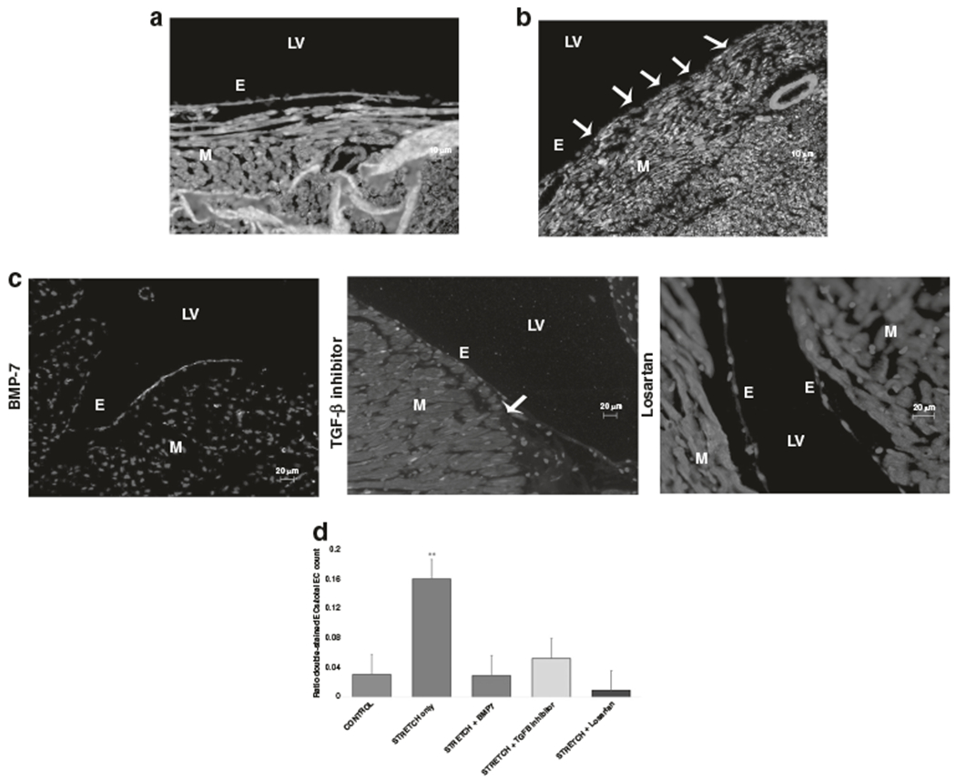
Fig. 3. Excess strain induces EndMT in immature hearts.

Heart tissue was stained for the endothelial marker PECAM1 (green), the mesenchymal marker αSMA (red), and DAPI (blue). Double staining for both PECAM1 and αSMA indicated EndMT (white arrows point out EndMT positive endothelial cells). Representative immunohistochemical stains for double-labeling of endothelial cells with PECAM1 and αSMA are shown for a control hearts, b immature rat hearts stretched for 3 h, c immature hearts stretched with an inhibitor present (BMP-7 on the left, TGF-β-inhibitor in the middle, Losartan on the right). d A summary of the results is shown in this graph. Ratio of double-stained (PECAM1/alpha-SMA) endothelial cells/total endothelial cell count in controls, stretched and stretch with inhibitors are shown. There were significantly more double-stained cells in stretched hearts compared to all other groups (**p < 0.05) but there was no significant difference between the other groups.
