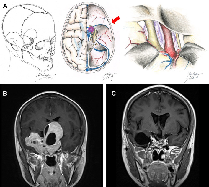
Figure 2.
Schematic diagram and MRI images of the frontotemporal approach. Schematic diagrams of incision, surgical field, and microanatomy of frontolateral approach (A). Preoperative (B) and 3-month postoperative (C) coronal enhanced MRI images of a giant pituitary adenoma in Knosp grade 4 that underwent the frontotemporal approach.

