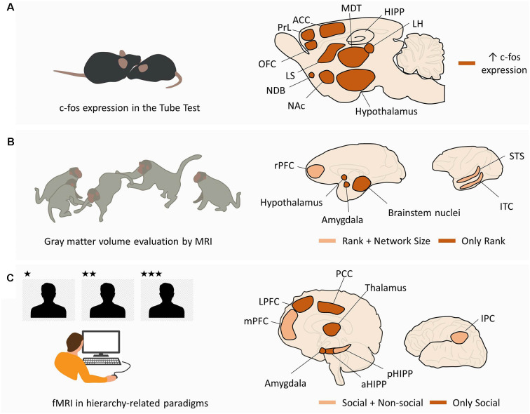
Figure 2.
Macroscale networks supporting social hierarchies. (A) Macroscale network in rodents. In mice performing tube tests for social hierarchy, c-fos expression in dominant animals is increased in the ventromedial hypothalamus, LH, ACC, medial preoptic area (hypothalamus), and MDT. In addition, pairwise correlations were found between MDT-PrL, MDT-OFC, MDT-ACC, MDT-CA2, ACC-MDT, ACC-LH, ACC-NDB, ACC-LS, and ACC-NAc. (B) Macroscale network in non-human primates. MRI studies in non-human primates from different social statuses and living in groups of different sizes have shown a correlation between social rank and gray matter volume in the hypothalamus, amygdale, and brainstem nuclei (“Only Rank”). Gray matter volume in rPFC, STS, and ITC was correlated with both social rank and group size (“Rank + Network Size”). (C) Macroscale network in humans. fMRI studies in human subjects performing hierarchy-related tasks have shown selective activation of LPFC, amygdala, aHIPP, thalamus, and PCC by the social component of the task (“Only Social”). The mPFC, pHIPP, and IPC were activated in the social and non-social conditions, suggesting a domain-general function (“Social + Non-social”). See text for other domain-general regions in human literature not depicted in the figure. Abbreviations: LH, lateral habenula; ACC, anterior cingulate cortex; MDT, medial preoptic area (hypothalamus), and mediodorsal thalamus; PrL, prelimbic cortex; OFC, orbitofrontal cortex; NDB, nucleus of the diagonal band; LS, lateral septum; NAc, nucleus accumbens; rPFC, rostral prefrontal cortex; STS, superior temporal sulcus; ITC, inferior temporal cortex; LPFC, lateral prefrontal cortex; aHIPP, anterior hippocampus; pHIPP, posterior hippocampus; PCC, posterior cingulate cortex; mPFC, medial prefrontal cortex; IPC, inferior parietal cortex.

