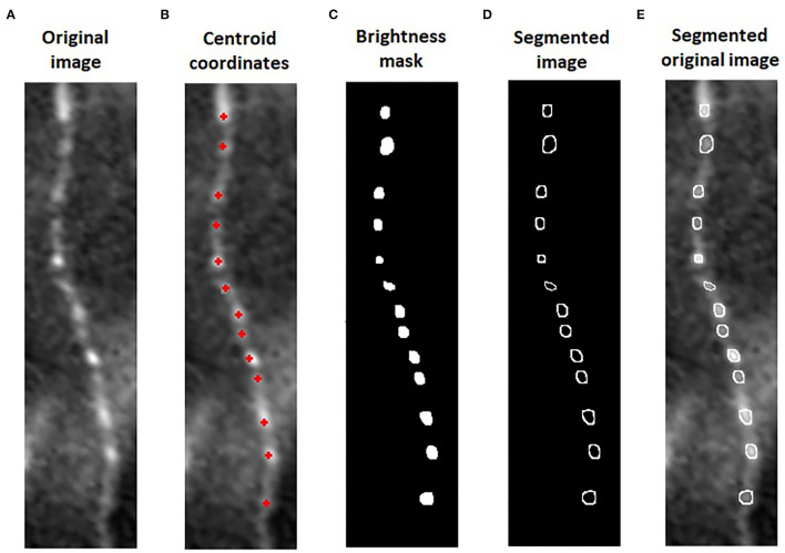
Figure 2.
Algorithm for automatic beading size (BS) measure: We cropped 0.1 mm of nerve in the original image of sub-basal plexus (A). The algorithm used the coordinates of corneal beadings' centroids from the image with overlaying analysis (B) to segment the area and defined the beadings in the original image. The intensity of each centroid was used to mask all the pixels below this value (C). On the original image, the perimeter of the beadings was segmented and the BS quantified (D,E).

