FIGURE 3.
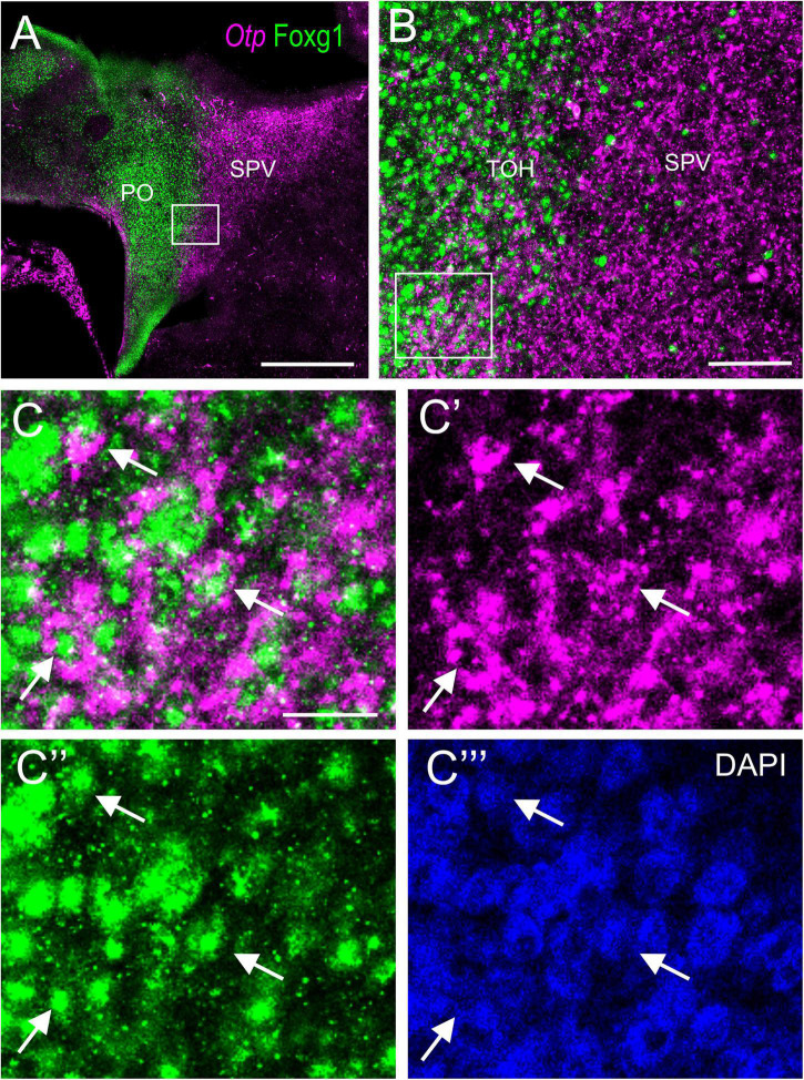
Fluorescent double labeling of Otp and Fogx1 in chicken. (A) Sagittal section of the chicken embryonic forebrain, at E8, hybridized for Otp (magenta color) and immunostained for Foxg1 (green color). Note the overlapping expression of both transcription factors in the TOH domain, just dorsal to the SPV core. (B) Detail of the area squared in (A) showing coexpression of Otp and Foxg1 in many cells of the TOH domain. Panels (C–C′′′) show the area squared in B at higher magnification (C: merged magenta/green channels; C′,C′′: separate magenta and green channels; C′′′: DAPI staining). Arrows point to a few examples of double-labeled cells. See text for more details. For abbreviations, see list. Scale bars: (A) = 320 μm; (B) = 80 μm; (C) = 20 μm (applies to C–C′′′).

