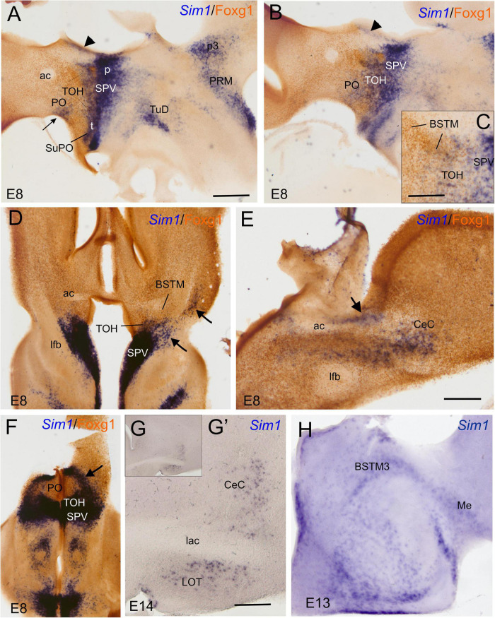
FIGURE 8.
Chromogenic labeling of Sim1 and Foxg1 in the chicken forebrain. (A–C) Sagittal, (D,F) horizontal, and (E) frontal sections of the chicken embryonic forebrain, at E8, hybridized for Sim1 (blue color) and immunostained for Foxg1 (brown color). Note the overlapping expression of both transcription factors in the TOH domain, just dorsal to the SPV core. Panel (C) shows a detail of the overlapping area covering part of BSTM (from a section a bit lateral to that seen in B). Arrowheads in (A,B) show a stream of Sim1 cells spreading dorsally from the SPV core. This stream reaches the BST (pointed with an arrow in E). A lateral stream of Sim1 cells (arrow in panel D) also reaches the capsular central amygdala and adjacent arcopallium (E). The arrows in (D,F) point to streams of Sim1 expressing cells spreading from the TOH and SPV domains to the capsular (D) central amygdala or (F) the PO. Panels (G–H) show details of frontal sections hybridized for Sim1 at the level of the (G,G′) central or the (H) medial extended amygdala in older embryos (E13 and E14). See text for more details. For abbreviations, see list. Scale bars: (A) = 400 μm (applies to A,B,D,F); (C,E) = 200 μm; (D) = 40 μm (applies to D,E). (G′) = 400 μm (applies to G′,H).

