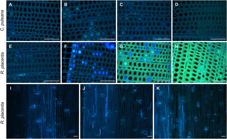
Figure 4.
Fluorescence images (ex: 330–385 nm, em: 420-nm) from decayed heartwood blocks. Transverse sections from C. puteana-exposed wood at sample positions 1 (A), 5 (B), 6 (C), and 7 (D), transverse sections from R. placenta-exposed wood at positions 1 (E), 3 (F), 4 (G), and 5 (H), and radial sections from R. placenta-exposed wood at positions 1 (I), 6 (J), and 7 (K). Images (A–H) were collected after 30 s of UV exposure in the microscope (330–385 nm), using the same acquisition settings for every image. Images (I–K) were collected without additional UV exposure. Scale bars are 0.1 mm.

