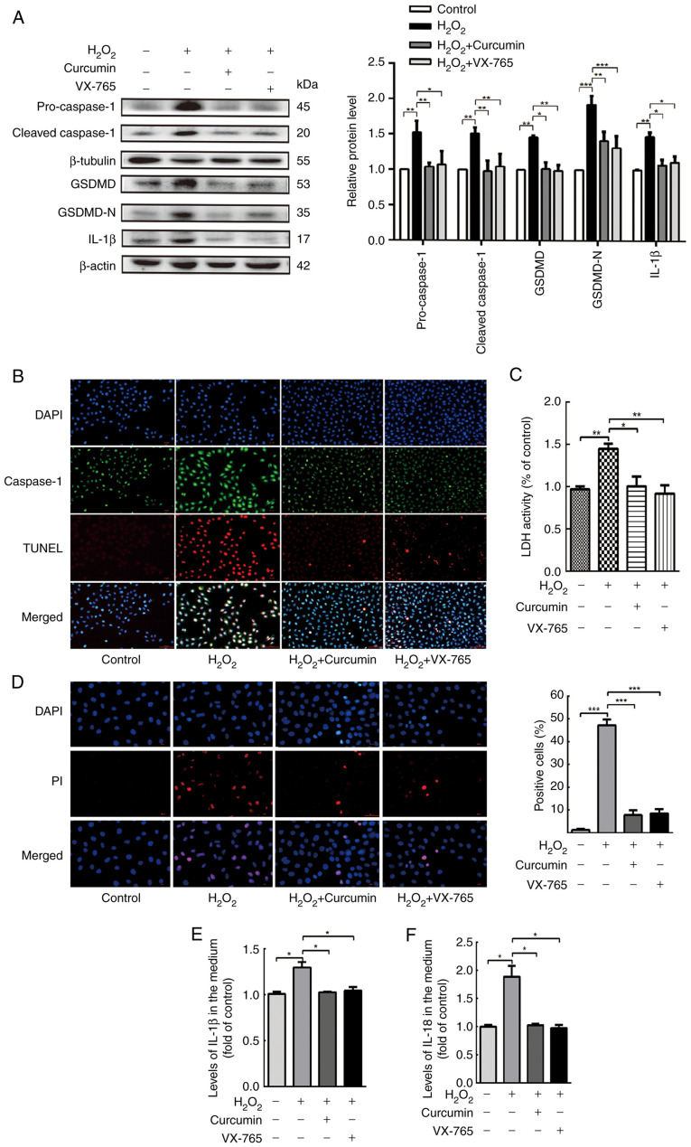
Figure 3.
Curcumin suppresses H2O2-induced pyroptosis of HUVECs. HUVECs were treated with H2O2 for 3 h and then either treated with 25 µM curcumin for 3 h or left untreated (control), whereas for the VX-765 group, HUVECs were pretreated with caspase-1 inhibitor VX-765 (10 µM) for 1 h and then incubated with H2O2 (800 µM) for 3 h. (A) Western blot analysis revealed that the protein levels of pro-caspase-1, caspase-1, pro-GSDMD, GSDMD and IL-1β were increased in HUVECs following treatment with H2O2 for 3 h. VX-765 and curcumin inhibited the protein expression of pro-caspase-1, caspase-1, pro-GSDMD, GSDMD and IL-1β. β-actin or β-tubulin was used as an internal control. (B) Compared with the H2O2 group, caspase-1 (green) and TUNEL (red) double-positive cells were decreased in the presence of curcumin or VX-765. The nuclei were stained blue with DAPI (magnification, ×400; scale bar, 50 µm). (C) The relative release of LDH in H2O2-treated HUVECs was increased, while that in curcumin- and VX-765-treated HUVECs was decreased (n=3). (D) The percentage of PI-positive HUVECs (red) was increased following H2O2 treatment, while it decreased following curcumin and VX-765 treatment (left, representative images; right, quantification of PI-positive cells; magnification, ×400; scale bar, 50 µm). Relative concentration of (E) IL-1β and (F) IL-18 in the culture medium following the treatment of HUVECs with H2O2, curcumin or VX-765, as determined by ELISA. Values are expressed as the mean ± standard deviation from three independent experiments. *P<0.05, **P<0.01 and ***P<0.001 as indicated. HUVECs, human umbilical vein endothelial cells; GSDMD, gasdermin D; LDH, lactate dehydrogenase; PI, propidium iodide.

