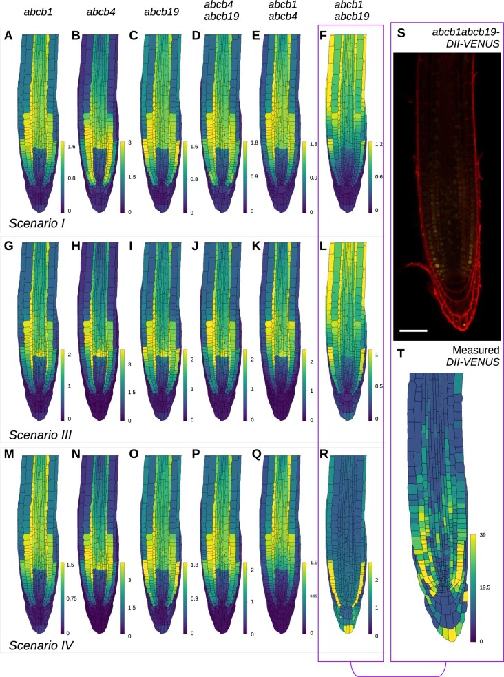
Figure 3.
Predicted DII-VENUS distributions for abcb single and double loss-of-function mutants for Scenarios I, III, and IV, together with experimental data showing the DII-VENUS distribution in abcb1 abcb19. A–R, Predicted DII-VENUS distribution for abcb1 (A, G, and M), abcb4 (B, H, and N), abcb19 (C, I, and O), abcb4 abcb19 (D, J, and P), abcb1 abcb4 (E, K, and Q), and abcb1 abcb19 (F, L, and R), in each of the three favored Scenarios I (A–F), III (G–L), and IV (M–R). S, Confocal image of abcb1 abcb19-DII-VENUS root-tip showing DII-VENUS (yellow) and cell geometries (red) (via propidium iodide staining). T, Measured DII-VENUS levels extracted from the image in (S).

