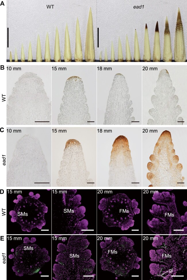
Figure 2.
ROS burst and PCD occur in ead1 apical ears. A, DAB-stained immature ears at 5, 7.5, 10, 12.5, 15, 17.5, 20, 22.5, 25, and 30-mm length in WT (left) and at 5, 7.5, 10, 12.5, 15, 17.5, 20, and >20-mm length in ead1 (right). Scale bars = 10 mm. B and C Paraffin sections of DAB-stained ear tips of WT (B) and ead1 (C) at different ELs. Scale bars = 100 μm. D and E TUNEL analysis of apical ears in WT (D) and ead1 (E). The first and third columns are from transverse sections and the second and fourth columns are from longitudinal sections. Sections were counterstained with propidium iodide. Signal was detected in ead1 (E), but not in the WT (D). gl, glume. Scale bars = 200 μm.

