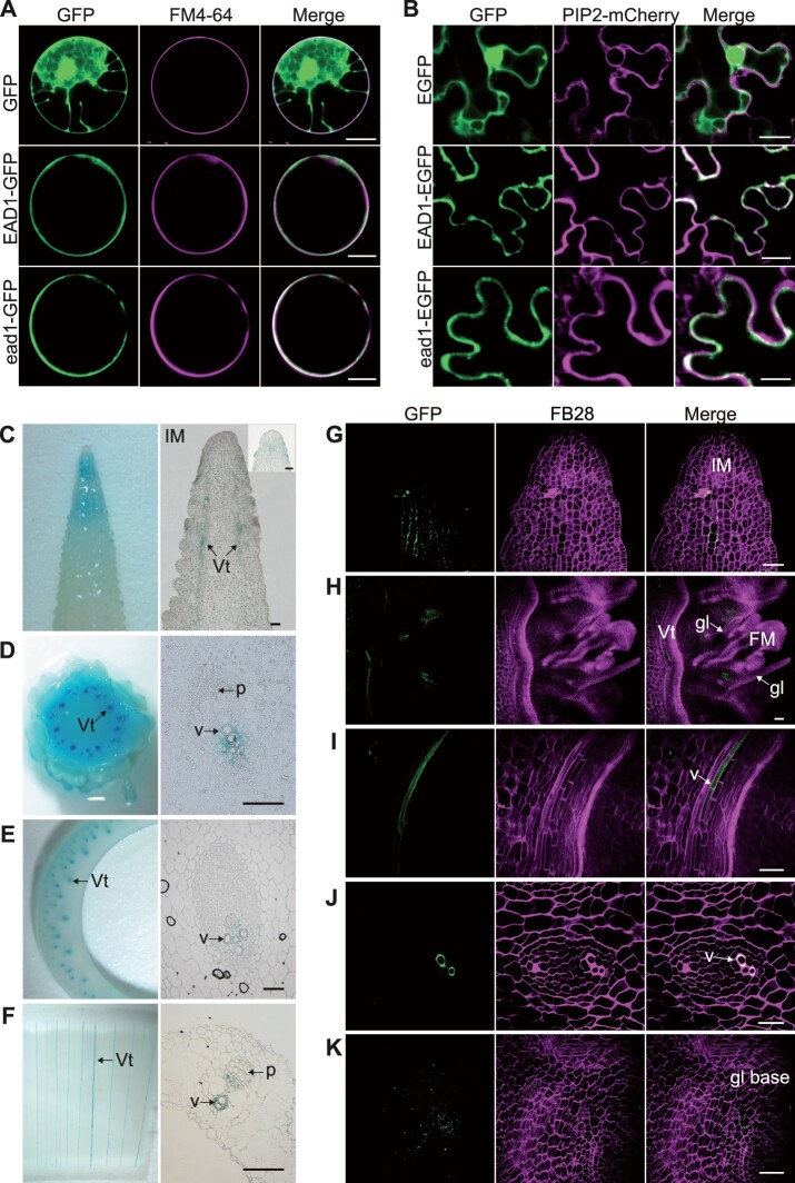
Figure 4.
Gene expression of EAD1 and protein tissue-specific localization of EAD1. A, Subcellular localization analysis in maize mesophyll protoplasts. The constructs Ubipro:GFP, Ubipro:EAD1-GFP, and Ubipro:ead1-GFP were transfected in maize protoplasts; the PM was stained with the dye FM4–64. Scale bars = 10 μm. B, Subcellular localization analysis in N. benthamiana epidermal cells. The constructs 35Spro:EGFP, 35Spro:EAD1–EGFP, and 35Spro:ead1–EGFP were separately co-infiltrated with the PM marker PIP2-mCherry. Scale bars = 20 μm. C–F, GUS staining results of ear related tissues in EAD1pro:GUS transgenic lines. C, Ear apical part at the 15-mm stage. D, Ear basal part at the 30-mm stage. E, Sheath of ear-leaf. F, Immature ear bract. Left: entire tissue; right, paraffin sections of GUS-stained tissues. Scale bars = 100 μm. G–K, Tissue-specific accumulation of the EAD1–GFP fusion protein in EAD1pro:EAD1–GFP transgenic maize ears. Positive signals were detected below the apical IM (G), the basal parts of FM glumes (H and K), and xylem vessels of the immature ears (H, I, and J). G, H, I, and K, the longitudinal sections and (J) indicates the transverse section from immature ears. K, the close-up view of the basal part of glume. Vt, vascular tissue; v, xylem vessel; p, phloem; gl, glume; BF, brightfield. FB28 is cell wall dye. Scale bars = 50 μm.

