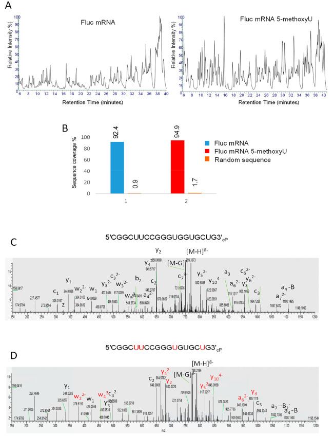
Figure 6.
RNA sequence mapping of chemically modified mRNA. (A) Total ion chromatograms of the partial RNase T1 digests of Fluc mRNA and Fluc 5-methoxyU mRNA. Twenty micrograms of RNA was incubated with 1.25 μL of immobilized RNase T1 for 10 min at 37 °C prior to LC–MS/MS analysis. (B) Bar chart showing the % sequence coverage of the partial RNase T1 digest of Fluc mRNA (ORF sequence), chemically modified mRNA (ORF), and a random RNA sequence. (C, D) MS/MS spectra of the oligoribonucleotide CGGCUUCCGGGUGGUGCUGcP and the corresponding oligoribonucleotide where the uridines are replaced with 5-methoxyuridines. The corresponding fragment ions are highlighted, and those fragment ions specific to the 5-methoxyuridine oligoribonucleotide are highlighted in red.

