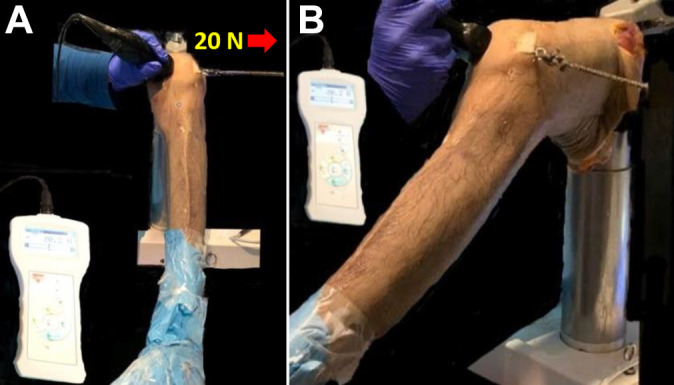
Figure 1.

Experimental setup demonstrating probe placement of the portable ultrasound on a left knee. (A) The probe is positioned at the medial patella at its widest portion and oriented parallel to the joint line to visualize the medial patellar facet and the medial trochlear facet on 1 image. Ultrasound images were obtained with and without 20 N of laterally directed force. (B) Setup shown from a different angle.
