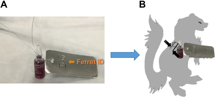
Figure 2.
A 1- to 2-inch incision was made between the ribs on either side of the thoracic vertebrae, approximately 2 to 3 inches below the neck area (opposite the chest/abdominal cavity area). Scalpels were used to cut into the flesh and through to the chest cavity, taking care to minimize the width of the incision hole. To implant the biological indicators (BIs) inside the abdominal cavity of the carcass, a flexible wire write-on metal tag (VWR, Radnor, PA) was wrapped around the BI (A), then the BI was fed through one of the incisions made on the back of the carcass and fixed into place by twisting the wire around the spine through the second incision hole (B).

