Figure 13.
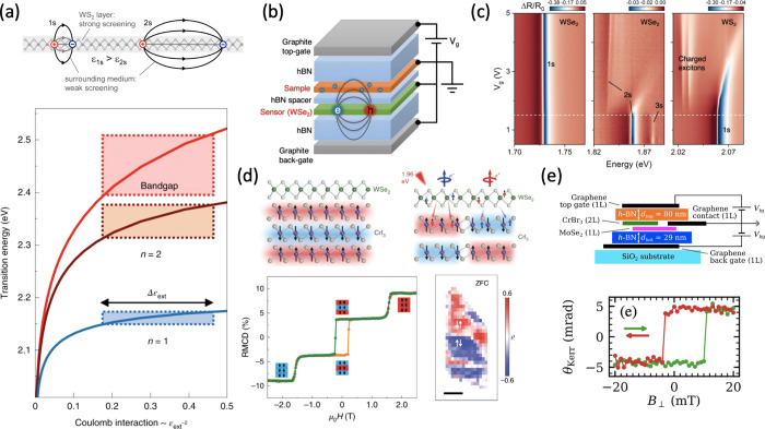
(a) Top: Schematic illustration of electron–hole pairs forming 1s and 2s excitonic states in a 2D dielectric slab. Adapted with permission from ref (151). Copyright 2014 American Physical Society. Bottom: Theoretically calculated energies of the bandgap and exciton states in a WS2 ML as a function of inverse squared external dielectric constant. Shaded areas indicate fluctuations from variations of the external screening. Adapted with permission from ref (202). Copyright 2019 Springer Nature. (b) Schematic of a device heterostructure to demonstrate the sensing capabilities of excitons in ML WSe2. (c) Reflectance contrast of the 1s (left) and 2s and 3s (middle) excitonic transitions in the WSe2 sensor, and the 1s exciton in the WS2 sample as a function of the applied gate voltage (i.e., the electron concentration in WS2). (b) and (c) Adapted with permission from ref (199). Copyright 2020 Springer Nature. (d) Left: Reflective magneto-circular dichroism as a function of magnetic field (bottom) measured in a monolayer WSe2 and trilayer CrI3 heterostructure, depicted above schematically. Orange and green curves represent magnetic field sweeping up (increase) and down (decrease), respectively. Right: Schematic of a ML WSe2 and bilayer CrI3 heterostructure (top). The layered AF spatial domains that are indistinguishable by reflective magneto-circular dichroism can be resolved by circular polarization-resolved PL from WSe2 (bottom). Adapted with permission from ref (203). Copyright 2020 Springer Nature. (e) Measurement of the CrBr3 magnetization hysteresis using the MOKE (bottom) in a device like schematically shown on top. Adapted with permission under a Creative Commons CC BY 4.0 license from ref (204). Copyright 2020 American Physical Society.

