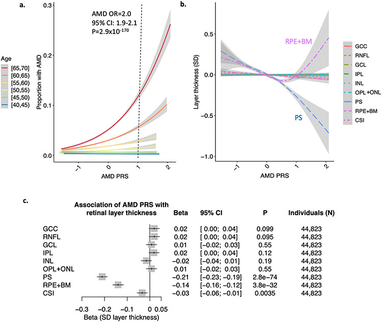
Figure 3: Associations of the AMD PRS with retinal layer thicknesses.
(a) association of AMD PRS with AMD (prevalence + incident AMD) by 5-year age categories at enrollment. A 1 unit increase in the AMD PRS confers a 2-fold increased risk of AMD (95%CI 1.9-2.1, P=2.9x10−170), after adjustment for sex, age, age2, smoking status, and principal components of genetic ancestry. Further adjusted associations by age category are provided in Supplementary Figure 7. (b) relationship of AMD PRS with layer thickness highlights strong negative correlation with the PS and a U-shaped relationship with the RPE layer. Curves and standard errors in panels (a) and (b) reflect the best-fit generalized additive model (gam) to the individual-level data, with added smoothness. (c) association of AMD PRS with retinal layer thickness, adjusted for sex, age, age2, smoking status, and principal components of genetic ancestry. GCC = ganglion cell complex, RNFL = retinal nerve fiber layer, GCL = ganglion cell layer, IPL = inner plexiform layer, INL = inner nuclear layer, OPL = outer plexiform layer, PS = photoreceptor segment layer, RPE+BM = retinal pigment epithelium plus Bruch’s membrane layer, CSI = choroid scleral interface.

