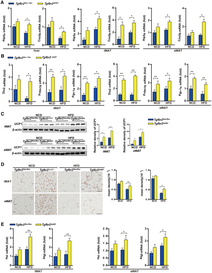
FIG. 3.

Ablation of Tgfbr2 in hepatocytes enhances mitochondrial oxidative phosphorylation and thermogenesis in mice fed an HFD. Tgfbr2flox/flox and Tgfbr2ΔHEP mice were fed an NCD or an HFD for 16 weeks; n = 7 per group. (A) mRNA expression of mitochondrial markers in the liver, iWAT, and eWAT. (B) The mRNA expression levels of browning genes in iWAT and eWAT. (C,D) UCP1 expression in iWAT and eWAT tissues by western blot and immunohistochemical staining (×400; black bar represents 5 μm) and the corresponding density of iWAT and eWAT tissues. (E) iWAT and eWAT lipolytic gene expression determined by real‐time PCR. Two‐way ANOVA was used for all statistical analysis. Data are represented as the mean ± SEM (*P < 0.05, **P < 0.01). Tgfbr2ΔHEP ‐NCD versus Tgfbr2flox/flox ‐NCD and Tgfbr2ΔHEP ‐HFD versus Tgfbr2flox/flox ‐HFD were examined.
