Figure 2:

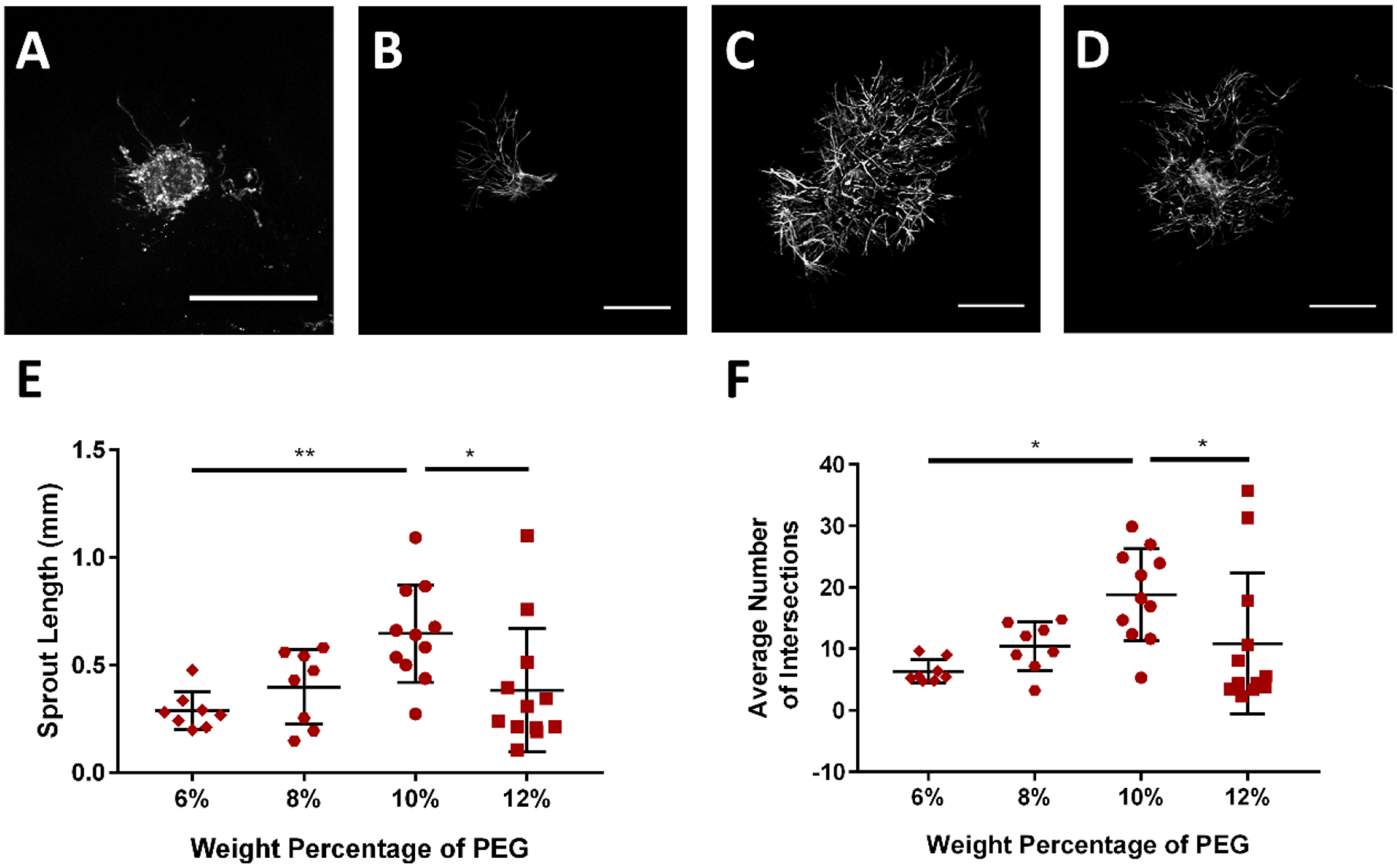
PEG hydrogel polymer weight percentage (WP) controls lymphatic sprouting. Representative images of collecting vessel segments at day 7 cultured in (A) 6% PEG, (B) 8% PEG, (C) 10% PEG, and (D) 12% PEG gels presenting 2.0 mM RGD. Images are of vessels labeled with Calcein-AM indicating live cells. Scale bar = 500 μm. (E) Sprout length and (F) average number of intersections exhibit maximal values at intermediate gel polymer densities. Each point represents a biologically independent sample (sample size: 6% PEG = 8, 8% PEG = 8, 10% PEG = 11, 12% PEG = 12). Data analyzed using Kruskal-Wallis test followed by Dunn’s multiple comparisons.
* p<0.05, ** p<0.01.
