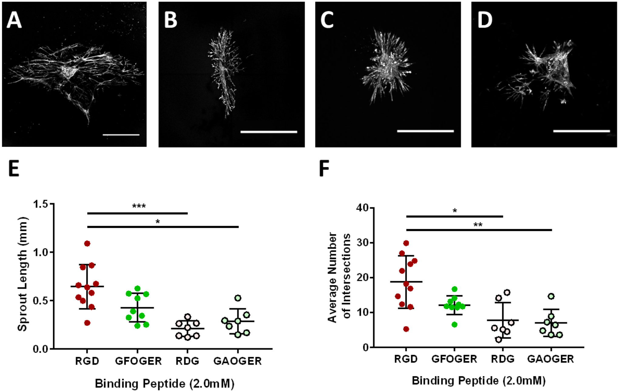
Figure 3:

RGD adhesive peptide promotes lymphatic sprout length and number in fully degradable PEG hydrogels. Representative images of collecting vessel segments stained with Calcein-AM at day 7 cultured in 10% PEG gels presenting (A) RGD, (B) GFOGER, (C) RDG, and (D) GAOGER peptides (2.0 mM). Scale bar = 500 μm. Quantification of (E) sprout length and (F) average number of intersections at day 7 compared to the GFOGER adhesive peptide and the scrambled control peptides RDG and GAOGER. Each point represents a biologically independent sample (sample size: RGD = 11, GFOGER = 9, RDG = 7, GAOGER = 7). Data analyzed using Kruskal-Wallis test followed by Dunn’s multiple comparisons. * p<0.05, ** p<0.01, *** p<0.001.
