Figure 6:

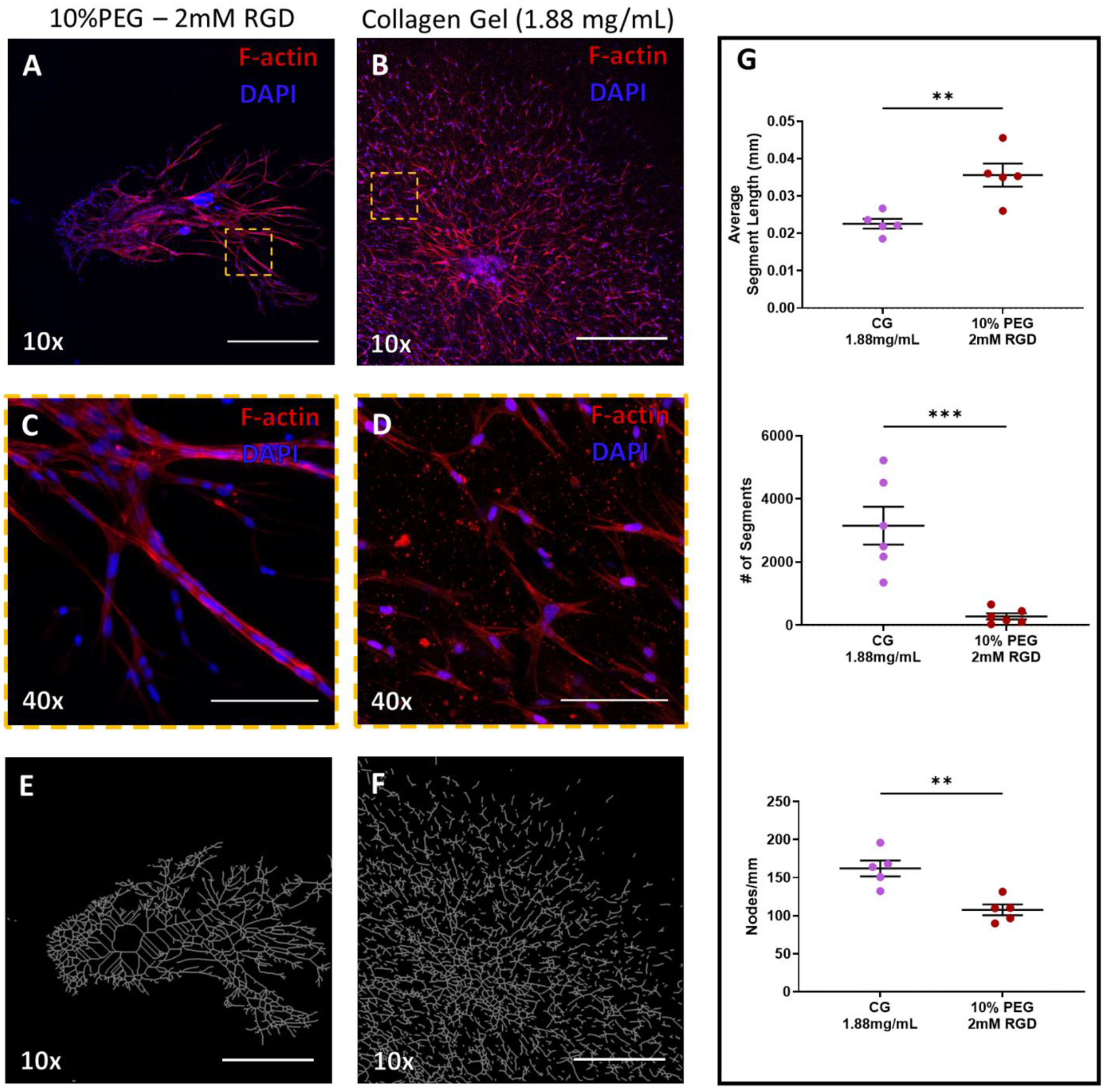
Culture of Lymphatic vessel segments in tunable PEG-4MAL hydrogels (A & B) F-actin (red) staining of lymphatic tissue at day 7 of culture in PEG hydrogel (fully degradable, 10% PEG − 2.0 mM RGD) or collagen gel (1.88 mg/mL), respectively. DAPI stain of nuclei in blue. Scale bars = 500 μm. (C & D) Indicated regions in panels (A) and (B), respectively, imaged with higher objective magnification demonstrates the varied organization of cells at 7 days of culture. Scale bar = 100 μm. (E & F) Simplified branching network produced by angiogenesis analyzer software based on F-actin staining in the PEG hydrogel (A) and collagen gel (B), respectively. (G) Sprouting metrics of average segment length, number of segments, and nodes/mm of PEG gel and collagen gel replicates.
