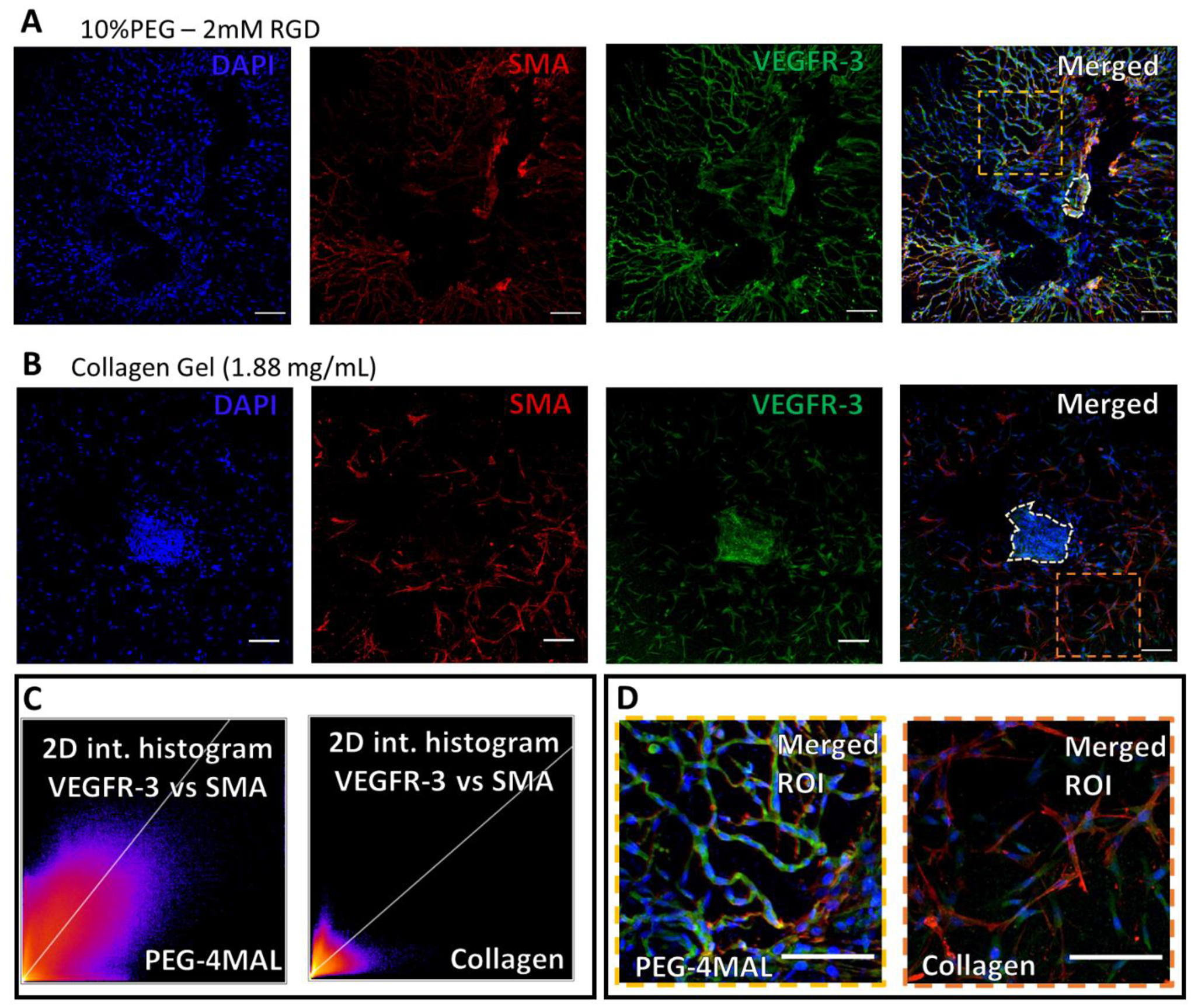
Figure 7: LEC and LMC distribution in lymphatic sprouting network after 7-day culture in hydrogels.

(A) Staining of vessel segments cultured in fully degradable, 10%PEG − 2 mM RGD hydrogels for nuclei (blue), α-smooth muscle actin (red), endothelial nitric oxide synthase (gray), and vascular endothelial growth factor receptor-3 (green), respectively. 2D intensity histograms demonstrating the correlation of VEGFR3 and SMA in the PEG-4MAL gel and collagen gel (B) Similar staining for vessel segments cultured in collagen gels (1.88 mg/mL). (I) Zoomed in and merged image of the vessel segment sprouting phenotype in PEG hydrogel. From region indicated in panel (A). (J) Zoomed in, merged, image of vessel segment sprouting phenotype in collagen gel (1.88 mg/mL). From region indicated in panel (E). Scale bars=100 μm.
