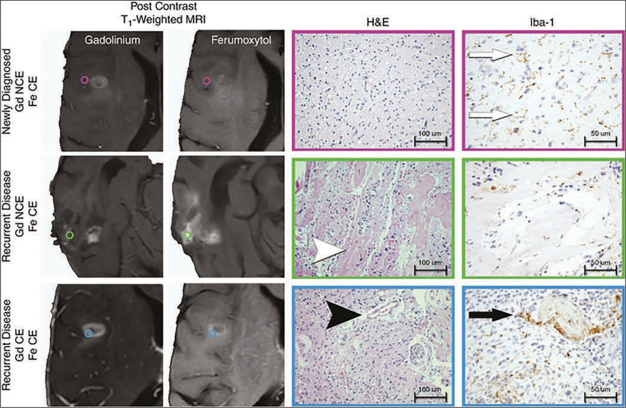
Fig. 7.

Histopathologic correlation of lesions enhancing after ferumoxytol (Fe)- and gadolinium (Gd)-based contrast agent administration in newly diagnosed and recurrent glioblastoma. Photomicrographs show regional image-guided tissue samples based on ferumoxytol and gadolinium contrast enhancement (CE) patterns. Standard-of-care stereotactic tissue sampling was performed in three patients with isocitrate dehydrogenase 1 (IDH1) wild-type glioblastoma at time of initial diagnosis or at time of disease recurrence. Tissue samples were classified by presence of gadolinium (left) or ferumoxytol (second from left) contrast enhancement. Tissue specimens were histopathologically characterized by presence of tumor and microvascular proliferation (H and E, ×20; 100-μm scale bar [second from right]) and presence of activated microglia or macrophages (ionized calcium-binding adapter molecule 1 [Iba1] stain, ×40; 50-μm scale bar [right]). Regions of ferumoxytol contrast enhancement but absence of gadolinium enhancement (NCE) were observed in patients with newly diagnosed glioblastoma (purple, circles, top row) and disease recurrence (green, circles, middle row). Ferumoxytol-only enhancing regions in patients with newly diagnosed IDH1 wild-type glioblastoma exhibited infiltrating glioma with low cellularity, delicate vasculature, and activated microglia (Iba1 brown-staining cells [white arrows]). Ferumoxytol-only enhancing regions in patients with recurrent IDH1 wild-type glioblastoma (blue, circles, bottom row) exhibited therapeutic changes evidenced by widespread vascular hyalinization (white arrowhead) with scattered macrophages without evidence of viable tumor. Dual contrast-enhancing sites (bottom row) appeared biologically similar in new diagnosis and disease recurrence settings, being characterized by highly cellular tumor with microvascular proliferation (black arrowhead) and tumor-associated macrophages with epithelioid appearance (black arrow). (Reprinted from [44] and in public domain)
