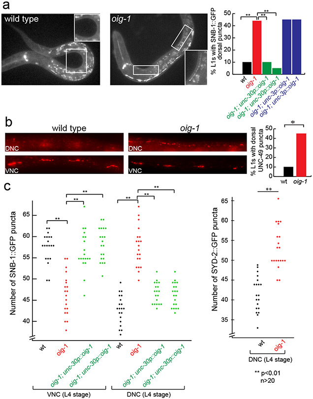
Figure 3: Aberrant D-type MN synapse formation in oig-1 mutants.
a: Ectopic DD synapses in the dorsal nerve cord of oig-1 mutant L1s.
b: Ectopic UNC-49 localization in oig-1 mutant L1s, as assessed by UNC-49 antibody staining. UNC-17 staining was used as a control to identify the ventral and dorsal nerve cords of the animals (not shown).
c: Ectopic VD synapses in the dorsal nerve cord of L4 staged oig-1 mutants. For each marker in each strain, puncta in the posterior of L4 animals from DD4 to VD11 were scored.

