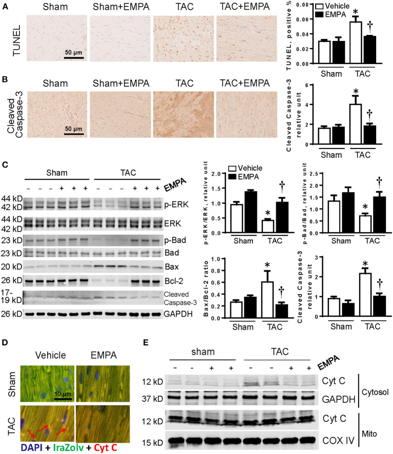
Figure 4.
EMPA treatment attenuated apoptosis in TAC-induced HF. (A) TUNEL staining of heart sections in sham, sham + EMPA, TAC and TAC + EMPA groups. (B) Immunohistochemistry results of cleaved caspase-3 in different groups. (C) Left, representative blots of proteins related to cellular survival and apoptotic pathways and Right, quantitative results. (D,E) Increased Cyt C released from mitochondria into cytoplasm after TAC is attenuated by EMPA treatment. Results are expressed as mean ± SEM, n = 5–7, *p < 0.05 vs. corresponding sham group, †p < 0.05 vs. corresponding TAC vehicle group. One-way ANOVA and Tukey post hoc test. EMPA, empagliflozin; SEM, standard error of the mean; TAC, transverse aortic constriction; ERK, extracellular signal-regulated kinase; Cyt C, cytochrome C; COX IV, cytochrome c oxidase, complex IV in the mitochondrial respiratory chain; DAPI, 4',6-diamidino-2-phenylindole; GAPDH, glyceraldehyde 3-phosphate dehydrogenase.

