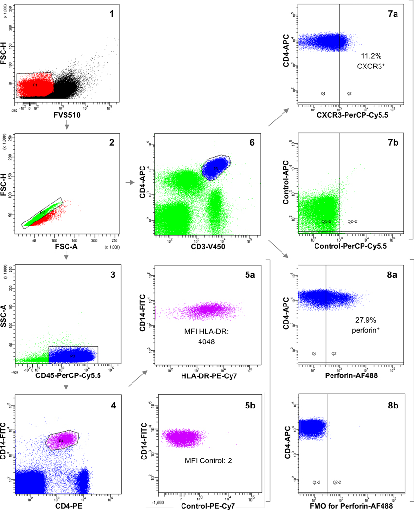
Figure 1.

Flow cytometry gating strategy. FVS510 was used to select viable cells (1). After gating for singlets (2), CD45+CD14+CD4+ cells (monocytes) were selected (3 and 4) and linear mean fluorescence intensity (MFI) values of HLA-DR were registered (5a). The absence of fluorescence for PE-Cy7 on monocytes was confirmed employing isotype control (5b). On the other hand, CD3+CD4+ cells (CD4+ T cells) were gated (6). The percentage of CD4+ T cells that were positive for CXCR3 (7a) and PD1 or negative for CD27 was assessed employing isotype controls (7b), whereas the percentage of CD4+ T cells that were positive for perforin (8a) and granzyme B was evaluated using fluorescence-minus-one (FMO) controls (8b). In the plots, fluorescence intensity values are transformed into logarithmic scale.
