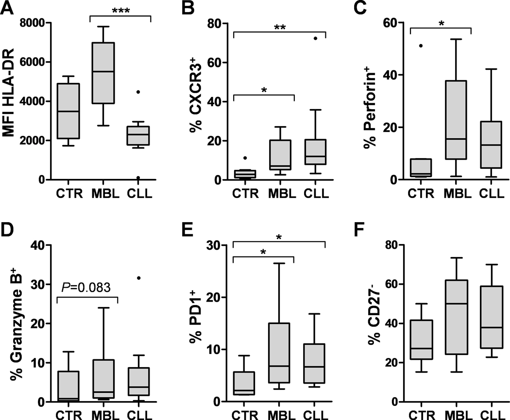
Figure 3.

Flow cytometry analysis. MFI of HLA-DR was assessed on monocytes (A), and the percentages (%) of CD4+ T cells that were positive for CXCR3 (B), perforin (C), granzyme B (D), PD1 (E) or negative for CD27 (F) were registered. Significant P-values are indicated with * (<0.05), ** (<0.01) or *** (<0.001). CTR: Healthy controls.
