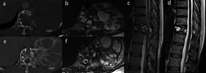
Fig. 5.
Image of a giant cell tumor of T11 in a 50-year-old woman. a, e Axial CT scans before and after denosumab treatment. Complete peripheral rim and bone formation inside the mass over 50% of the transverse session are observed (grade 3). b, f Axial MR images before and after denosumab treatment. The maximum diameter of the tumor and the proportion of the area of the spinal canal occupied by the tumors are smaller. c, d Sagittal MR images before and after denosumab treatment. The spinal cord compression completely disappears

