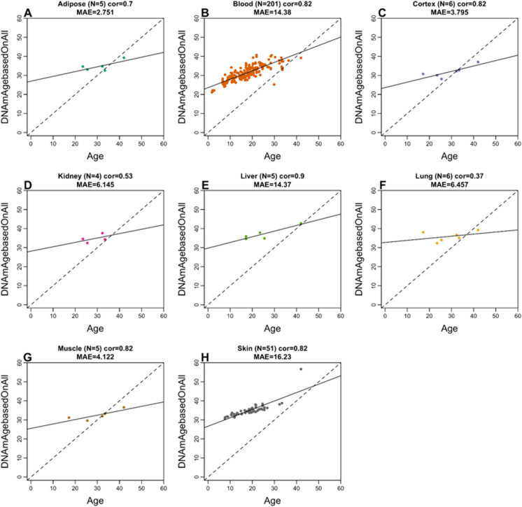
Fig. 3.
Multi-tissue vervet monkey clock applied to tissues from rhesus macaques. Each dot corresponds to a tissue sample from rhesus macaques: A adipose, B blood, C brain cortex, D kidney, E liver, F lung, G muscle, H skin. The y-axis reports the age estimate according to the multi-tissue vervet clocks. The predicted DNAm age in macaque tissues according to the vervet pan-clock (y-axis) and chronological age of the rhesus specimens (x-axis). The number of samples is shown in parentheses; cor, Pearson’s correlation; MAE, median absolute error

