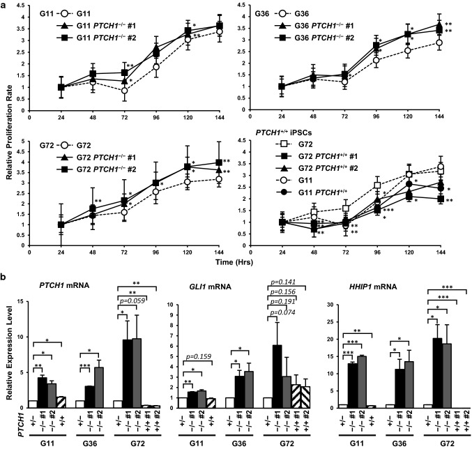
Fig. 2.
Hh signaling was accelerated in PTCH1−/− iPSC lines. a Cell proliferation assay of PTCH1-edited iPSCs. Eight hundred cells were plated on a 96-well plate and cell numbers were measured every 24 h using the BrdU incorporation assay. b The expression levels of the Hh-signaling target genes, PTCH1, GLI1, and HHIP1 were normalized by GAPDH mRNA levels. Data are presented as means ± SEM (n = 3). *: p < 0.05, **: p < 0.01, ***: p < 0.001

