
Fig. 5.
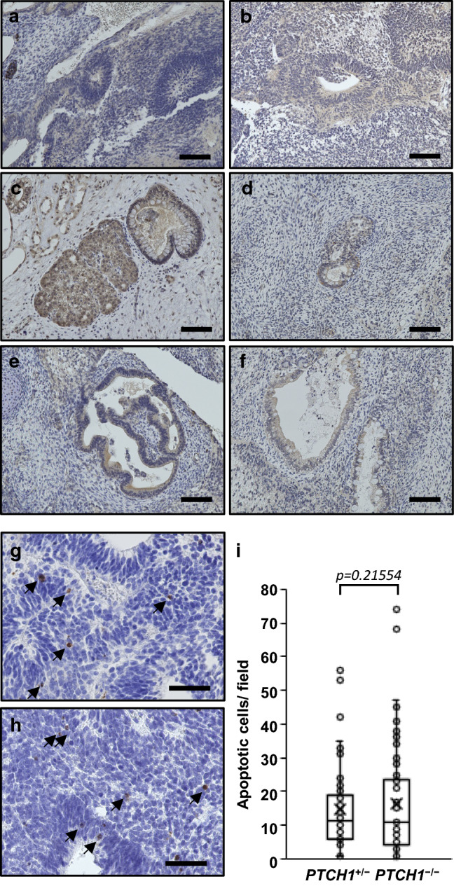
The SHH expressions and apoptotic cells in teratomas. a–f Immunohistological staining of SHH. SHH was expressed in neural tissues (a, b), glands c, d and intestinal epithelium e, f in PTCH1+/− a, c and e and PTCH1−/− teratomas (b, d and f). g–i Apoptosis in PTCH1+/− g and PTCH1−/− teratoma (h). Cleaved caspase-3 positive apoptotic cells were indicated by allows. Numbers of apoptotic cells counted in each field were displayed in the boxplot (i). Open circles and cross signs indicate each value of apoptotic cells and the averages, respectively (PTCH1+/−; n = 55, PTCH1−/−; n = 85). Scale bars: 100 μm in a–f, 50 μm in g and h