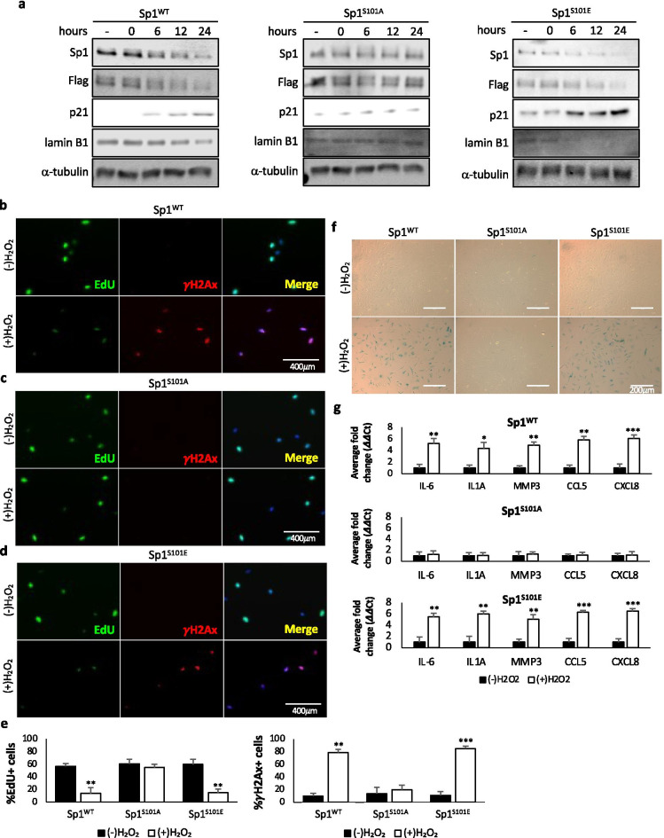
Fig. 3.
Phosphorylation of Sp1 by ATM is necessary for Sp1 degradation and damage-associated senescence. a–g hTert-BJ1 cells were depleted of Sp1 using CRIPSR/Cas9 and transduced with lentivirus expressing Flag-tagged Sp1WT, Sp1S101A, or Sp1S101E (Supplemental Fig. 1a). a Cells were treated with 200 μM H2O2 for 2 h, then placed in fresh media for 24 h. Lysates were collected at indicated time points past H2O2 release and used for Western blot analysis of protein levels. b–g Cells were treated with 200 μM H2O2 for 2 h, then placed in fresh media for 7 days. b–e 6 h prior to fixation, cells were treated with 10 μM EdU. Cells were then fixed and stained for EdU, γH2Ax, and DAPI. Scale bar represents 400 μm. f Cells described above were also stained for β-galactosidase. Scale bar represents 200 μm. g RNA was collected from cells described above. Quantitative RT-PCR was used to analyze samples with verified primers for SASP markers. GAPDH was used as a reference gene. Data were processed using the ΔΔCt method. Data represent means and SEM from 3 independent experiments assessed in triplicate. Significant differences between groups were determined by two-tailed Student’s t test. *, **, or *** indicate p values < 0.05, 0.01, or 0.001, respectively. No * indicates p value > 0.05

