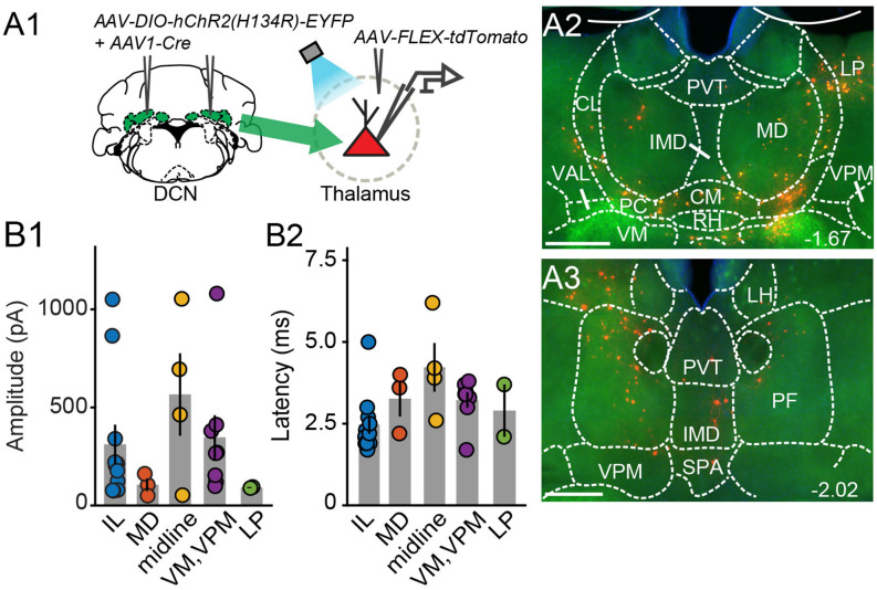
Figure 3.
Electrophysiologicalvalidation of virally-identified cerebello-thalamic connectivity. (A1) Schematic of experimental approach for ex vivo optophysiology. (A2,A3) Epifluorescence images of anterior (A2) and posterior (A3) thalamic slices acutely prepared for recordings. DCN input-receiving neurons are tdTomato+. Scale bars: 500 μm. (B) Average (± SEM) amplitude (B1) and onset latency (B2) of ChR2-evoked synaptic currents as a function of recording location in the thalamus. Intralaminar (IL) group: CL, PC, CM, and PF; midline group: IMD and RH.

