
FIGURE 1.
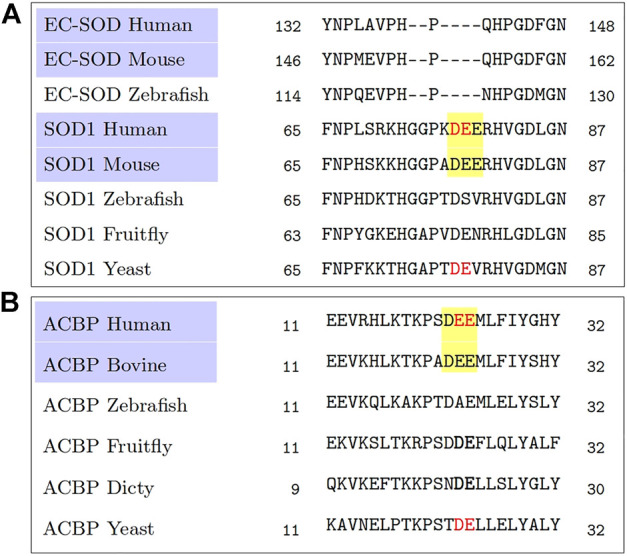
A reanalysis of the multiple sequence alignments obtained from Cruz-Garcia et al. (2017). A comparison among the mammalian sequences, highlighted in purple, shows a common triacidic motif DEE (highlighted in yellow), rather than a diacidic motif (shown in red colored text) when comparing sequences across all species. (A) Multiple sequence alignment from homologs of SOD1. (B) Multiple sequence alignment from homologs of ACB1.