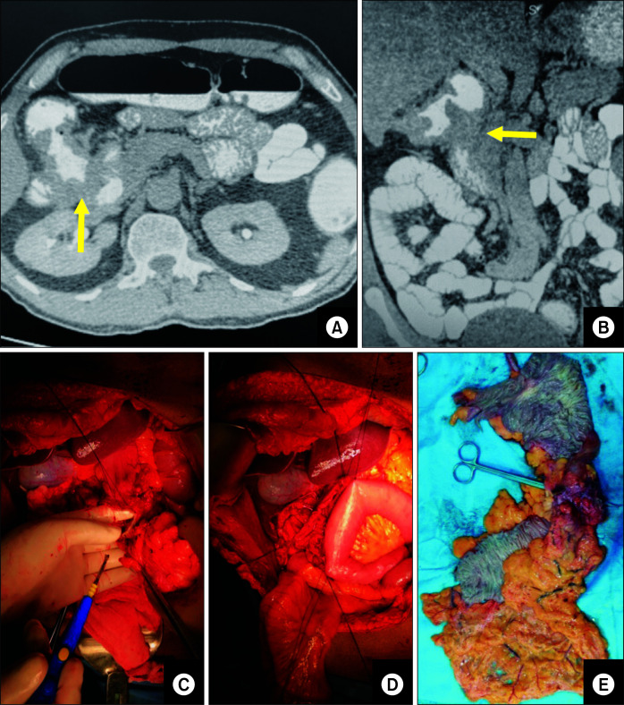
Fig. 4.
(A) Computed tomography (CT) image showing hepatic flexure growth with colo-duodenal fistula (arrow). (B) Coronal section of CT scan showing colo-duodenal fistula (arrow). (C) Intra-operative image showing duodenal involvement (specimen retracted by the first assistant). (D) Jejunal patch in progress (side-to-side loop). (E) Resected specimen showing hepatic flexure colonic growth with duodenal fistula.

