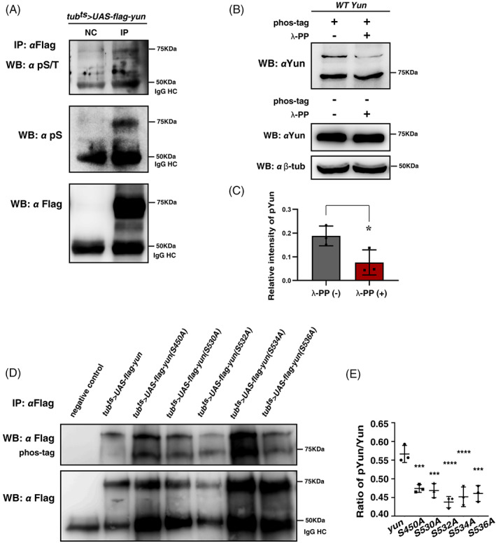
FIGURE 4.

Yun is phosphorylated in vivo. (A) Detection of the phosphorylation status of Yun (pYun) using anti‐phosphorylated serine/threonine (pS/T) and anti‐phosphorylated serine (pS) antibodies by IP and western blot, respectively. NC: negative control. The 50 KDa band is likely the IgG heavy chain (IgG HC). (B) Detection of pYun with phos‐tag and it can be dephosphorylated by λ‐PP. (C) Quantification of the grayscale of pYun with or without λ‐PP. Mean ± SD is shown. n = 3. *p < 0.05. (D) Detection of pYun with phos‐tag in flies expressing different Flag‐tagged Yun forms by tub ts . (E) Quantification of pYun/total Yun ratio of different Yun forms. Mean ± SD is shown. n = 3. ***p < 0.001; ****p < 0.0001
