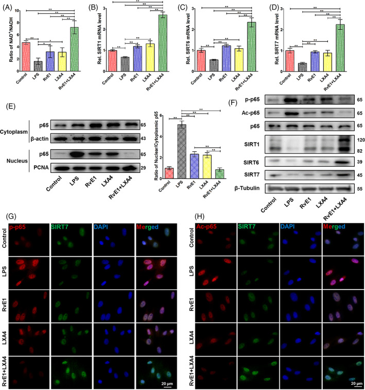
FIGURE 2.

Effects of the RvE1 and LXA4 combination on NF‐κB modification. (A) NAD+/NADH ratio measured by a NAD+/NADH assay kit. (B) SIRT1, (C) SIRT6, and (D) SIRT7 mRNA levels detected by qPCR. (E) Expression of p65 protein in both cytoplasm and nucleus detected by western blotting, normalized to β‐actin (in cytoplasm) or PCNA (in nucleus). (F) Phosphorylation and acetylation levels of p65 and expressions of SIRT1, SIRT6 and SIRT7 protein (normalized to that of β‐tubulin) detected by western blotting. (G) Representative double‐immunofluorescence labelling images of SIRT7 (green) and p‐p65 (red), and the nuclei were stained with DAPI (blue). (H) Representative double‐immunofluorescence labelling images of SIRT7 (green) and Ac‐p65 (red), and the nuclei were stained with DAPI (blue). (*p < 0.05 and **p < 0.01). DAPI, 4′,6‐diamidino‐2‐phenylindole; LXA4, lipoxin A4; NAD+, nicotinamide adenine dinucleotide; NF‐κB, nuclear factor kappa B; qPCR, quantitative polymerase chain reaction; RvE1, resolvin E1
