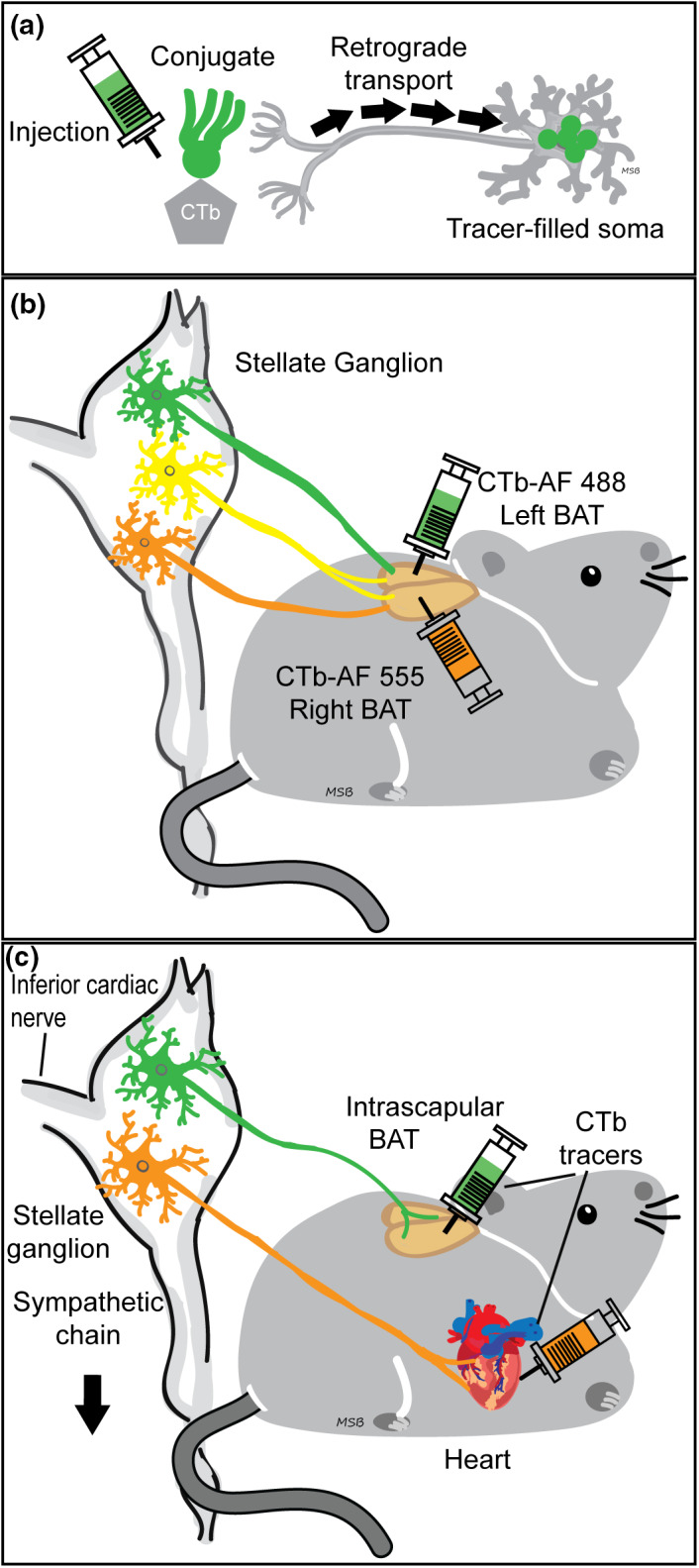
FIGURE 1.

Experimental design: Dual tracing methods retrogradely labeled different sympathetic neuron stellate ganglia (SG) subpopulations based on the tissue injected. (a): Schematic showing cholera toxin subunit b (CTb, Invitrogen) retrograde tracer uptake from axon terminals near injection site to somata, where the fluorescent conjugate (green) was detected. (b): Left and right intrascapular brown adipose tissue (BAT) dual tracer injections retrogradely labeled SG BAT‐projecting neurons. Using different tracer conjugates on each side allowed us to study the circuitry of the BAT‐projecting SG population, including bilateral projections (yellow). We also examined tracer viability at multiple post‐injection times (7, 14, and 28 days). (c): Dual tracer injection techniques allowed for direct comparison and quantification of two neuronal subgroups within SG: Intrascapular BAT projecting (green) and cardiac projecting (orange).
